ANP 1105 Lecture Notes - Lecture 4: Afterload, Stroke Volume, Ventricular Fibrillation

22 Mar 2016
School
Department
Course
Professor
Document Summary
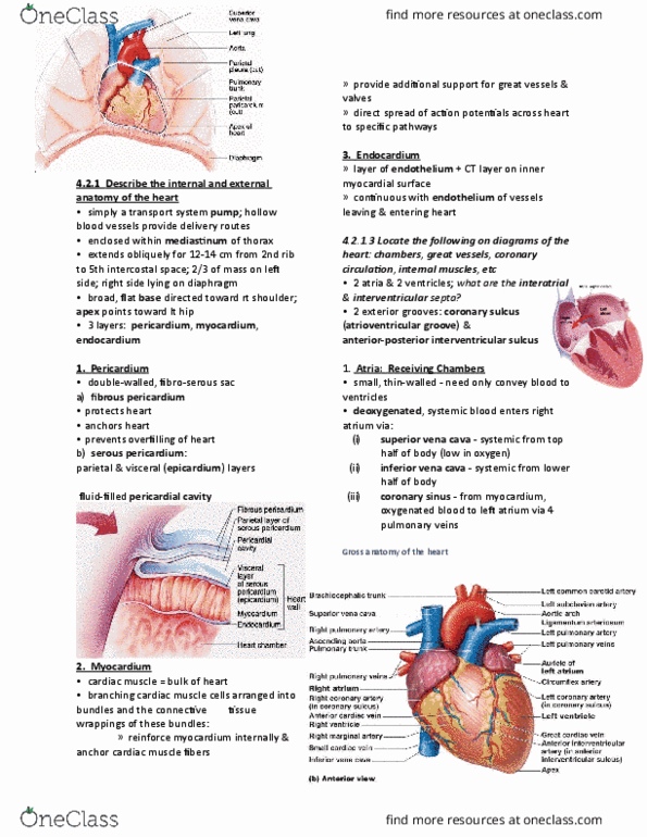
4. 2. 1 describe the internal and external anatomy of the heart. ~ fibrous pericardium protects and anchors heart: prevents overfilling. ~ serous pericardium: parietial and visceral epicardium layers: myocardium: cardium muscle = bulk of the heart. ~ branching cardiac muscle cells arranged into bundles and the ct that wraps around these muscles. The atria are the receiving chambers of the heart. They are small, thin walled, and their main task is to convey blood to the ventricles. Deoxygenated blood enters the right atrium via the superior and inferior vena cavas, as well as the coronary sinus; oxygenated blood travels to the left trium via 4 pulmonary veins. The ventricles are the pumps of the heart and have walls much thicker than the atria. The right ventricle pumps blood into the pulmonary trunk and the left ventricle pumps blood to the aorta. The nternal walls of the cventricles have muscle bundles called trabeculae carneae and papillary muscles.