NURS 1750 Lecture Notes - Lecture 5: Teres Minor Muscle, Abdominal Wall, Intercostal Nerves

28 Jun 2018
School
Department
Course
Professor

Week Five (Oct. 16-20, 2017)
Anatomy and Physiology
Chapter Thirteen: The Spinal Cord and Spinal Nerves (Pg. 446 - 474)
13.1 Spinal Cord Anatomy
- The spinal cord and spinal nerves contribute to homeostasis by providing quick, reflexive
responses to many stimuli. The spinal cord is the pathway for sensory input to the brain and
motor output from the brain.
- About 100 million neurons and even more neuroglia compose the spinal cord, the part of the
central nervous system that extends from the brain.
Protective Structures
Recall that nervous tissue of the CNS does not respond well to injury/damage.
Vertebral Column
- The first layer of protection for the CNS is the hard vertebral column.
- The skull encases the brain and the vertebral column surrounds the spinal cord,
providing strong protective defenses against damaging blows or bumps.
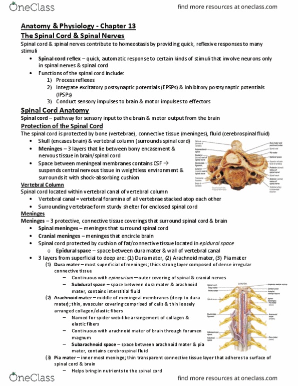
Meninges
- Three membrane that lie between the bony encasement and the nervous tissue
in both the brain and the spinal cord.
- Dura Mater: The most superficial of the three spinal meninges is a thick
strong layer composed of dense irregular connective tissue.
- Arachnoid Mater: The middle of the meningeal membranes, is a thin,
avascular covering comprised of cells and thin, loosely, arranged collagen
and elastic fibers.
→ It is called the arachnoid mater because of its spider’s web
arrangement of delicate collagen fibers and some elastic fibers.
- Pia Mater: The innermost meninx is a thin transparent connective tissue
layer that adheres to the surface of the spinal cord and brain. Consists of
thin squamous to cuboidal cells within interlacing bundles of collagen
fibers and some fine elastic fibers. There’s many blood vessels within pia
mater that supply oxygen and nutrients to the spinal cord.
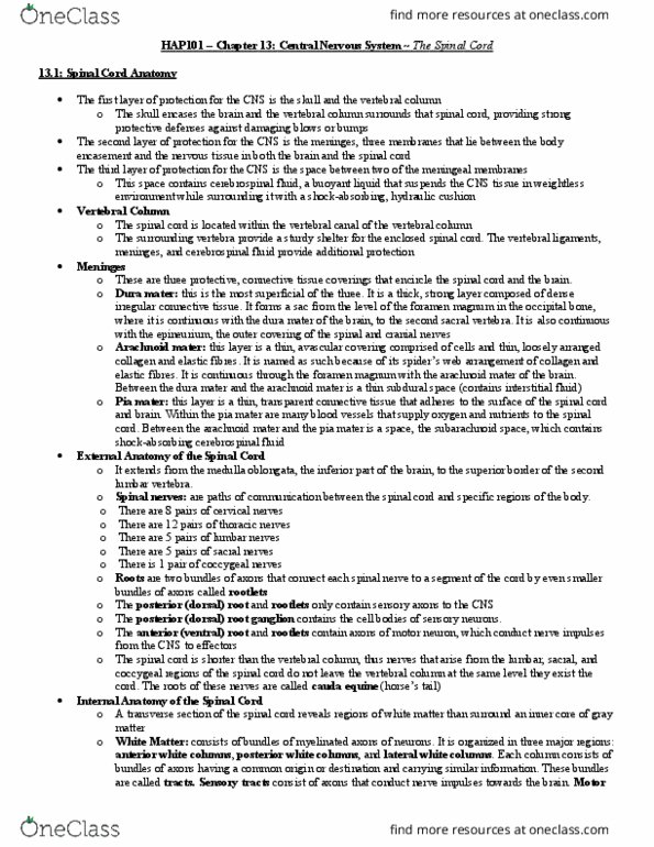
External Anatomy of the Spinal Cord
- In adults, the spinal cord extends from the medulla oblongata, the inferior part of the
brain, to the superior border of the second lumbar vertebra.
- In infants, the spinal cord extends to the third or fourth lumbar vertebra.
- When the spinal cord is viewed externally, two conspicuous enlargements can be seen:
- The Cervical Enlargement: extends from the fourth cervical vertebra (C4) to the
first thoracic vertebra (T1)
Internal Anatomy of the Spinal Cord
13.2 Spinal Nerves
find more resources at oneclass.com
find more resources at oneclass.com
Document Summary
Chapter thirteen: the spinal cord and spinal nerves (pg. The spinal cord and spinal nerves contribute to homeostasis by providing quick, reflexive responses to many stimuli. The spinal cord is the pathway for sensory input to the brain and motor output from the brain. About 100 million neurons and even more neuroglia compose the spinal cord, the part of the central nervous system that extends from the brain. Recall that nervous tissue of the cns does not respond well to injury/damage. The first layer of protection for the cns is the hard vertebral column. The skull encases the brain and the vertebral column surrounds the spinal cord, providing strong protective defenses against damaging blows or bumps. Three membrane that lie between the bony encasement and the nervous tissue in both the brain and the spinal cord. Dura mater: the most superficial of the three spinal meninges is a thick strong layer composed of dense irregular connective tissue.