NURS 1750 Lecture Notes - Lecture 2: Sodium Chloride, Trachea, Dna Replication

28 Jun 2018
School
Department
Course
Professor

Week Two (Sept. 18-22, 2017)
Anatomy and Physiology I
Chapter Three: The Cellular Level of Organization (Pgs. 61-102)
Cells and Homeostasis
Cells:
- Help each system contribute to the homeostasis of the body.
- All cells share key structures and functions that support their activity.
- Cells are basic, living, structural, functional units of the body.
- An average adult has 100 trillion cells.
- The scientific study of cells is called cell biology or “cytology”.
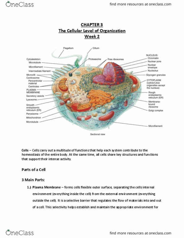
3.1 Parts of a Cell
Three Main Parts of a Cell:
- Plasma Membrane: flexible outer surface. Contains various proteins.
→ Separates cell’s internal environment from the external environment.
→ Regulates the flow of materials in/out of cell.
→ Participates in intercellular signaling.
- Cytoplasm: all the cellular contents between the plasma membrane and the
nucleus. Consists/contains cytosol (a.k.a intercellular fluid) and organelles
→ Cytosol: fluid portion of cytoplasm; contains water, dissolved solutes,
and suspended particles.
→ Organelles: each have a characteristic shape and specific function.
- Nucleus: a large organelle that contains most of the cell’s DNA.
Recall: a single molecule of DNA (called a chromosome) contains genes
that control cellular structure and function.
3.3 The Plasma Membrane
The plasma membrane is a is a structure that surrounds and contains the cytoplasm of
the cell. It is best described by using the fluid mosaic model → a model that shows the
plasma membrane as a moving sea of lipids containing many different proteins (mosaic).
Structure of the Plasma Membrane
The Lipid Bilayer
The lipid bilayer (framework of plasma membrane) is made up of two back-to-
back layers of phospholipids, glycolipids and cholesterol (all lipids).
→ The bilayer arrangement occurs because the lipids are amphipathic
molecules; meaning they have both polar and nonpolar parts
(phospholipids head’s are polar and tails are nonpolar).
→ Cholesterol molecules are weakly amphipathic. The polar -OH group
on cholesterols form H-bonds with the polar heads of phospholipids and
glycolipid while their steroid nonpolar rings fit among the fatty-acid tails of
the phospholipids and glycolipids.
find more resources at oneclass.com
find more resources at oneclass.com

→ Glycolipids have a carbohydrate group which forms a polar “head”.
Glycolipids appear only in the membrane layer that faces the extracellular
fluid; one reason the sides of the bilayer are different.
Arrangement of Membrane Proteins
Integral Proteins: are proteins used for transport. They are firmly embedded into
bilayer. Most integral proteins are transmembrane proteins; meaning they span
the entire bilayer, protruding into both cytosol and extracellular fluid.
→Though, a few integral proteins are tightly bound to one side of the
bilayer by covalent bonding to fatty acids.
Peripheral Proteins: are structural proteins. They are not as firmly embedded into
the bilayer. They are attached to the polar heads of lipids or to integral proteins.
Glycoproteins: are proteins with carbohydrate groups attached to the ends that
protrude into the extracellular fluid.
Glycocalyx: The sugary coat of the carbohydrate portion of glycoproteins and
glycolipids. The pattern of carbohydrates in the glycocalyx varies from cell to cell;
thus acting as molecular “signature” allowing cells to recognize each other.
Functions of Membrane Proteins
find more resources at oneclass.com
find more resources at oneclass.com

Week Two (Sept. 18-22, 2017)
Ion Channel: holes that specific ions (ie. K+) can flow through to get in/out of cell.
Usually selective to a single type of ion. Opening for specific ion.
Carrier (transporters): selectively moving a polar substance or ion from one side
to the other side of membrane. Substance to be transported.
Receptor (integral): serve as cellular recognition sites. Binds to specific type of
molecule. (ie. insulin receptors only bind to the hormone insulin). → A specific
molecule that binds to a receptor site is called a ligand. Binding site for function
of a cell.
Enzyme (integral and peripheral): catalyze specific chemical reactions at the
inside or outside surface of cell. Catabolizes reactions.Enzyme binds to lactose
and produces lactase.
→ ie. lactase protruding from epithelial cells lining your small intestine
splits the disaccharide lactose from milk.
Linker (integral and peripheral): they anchor proteins in the plasma membranes
of neighbouring cells to one another or to protein filaments inside and outside of
plasma membrane thus providing structural stability and shape for cell. Anchors
filaments for structure and attaches cells together.
Cell Identity Marker: Distinguishes your cells from anyone else’s. They can
recognize and respond to potentially dangerous foreign cells. Distinguishes your
cell from anyone else’s (except your twin).
→ An important class of such markers are the major histocompatibility
(MHC) proteins.
Membrane Fluidity
Membrane lipids and membrane proteins move easily in their own half of the bilayer.
Cholesterol: stabilizes the membrane and reduces membrane fluidity (keep
shape) in normal body temperatures.
○ At lower temperatures, cholesterol molecules breakup the packaging that
occurs as phospholipids solidify into gel → increasing the space between
tightly packed phospholipids and thus increasing fluidity. Fatty acids are
compressed in low temperatures; membrane becomes dense.
○ At high temperatures and room temperature, the presence of cholesterol
increases and the intramolecular forces in the membrane and holds
phospholipids more tightly together, reducing fluidity. Unsaturated fats
have kinks to keep phospholipids apart.
Factor Affecting Fluidity of the Plasma Membrane:
● Temperature:
find more resources at oneclass.com
find more resources at oneclass.com
Document Summary
Chapter three: the cellular level of organization (pgs. Help each system contribute to the homeostasis of the body. All cells share key structures and functions that support their activity. Cells are basic, living, structural, functional units of the body. An average adult has 100 trillion cells. The scientific study of cells is called cell biology or cytology . Separates cell"s internal environment from the external environment. Regulates the flow of materials in/out of cell. Cytoplasm: all the cellular contents between the plasma membrane and the nucleus. Cytosol: fluid portion of cytoplasm; contains water, dissolved solutes, and suspended particles. Organelles: each have a characteristic shape and specific function. Nucleus: a large organelle that contains most of the cell"s dna. Recall: a single molecule of dna (called a chromosome) contains genes that control cellular structure and function. The plasma membrane is a is a structure that surrounds and contains the cytoplasm of the cell.