Anatomy and Cell Biology 3319 Lecture Notes - Lecture 36: Hyaline Cartilage, Respiratory Tract, Trachea

1 May 2018
School
Department
Professor

Lecture 036: Trachea, Pleura, Lungs
Today’s Objectives
● Describe the passageway of air through the respiratory system
● Identify and describe the anatomical features of the respiratory system and their
relationships
● Describe the respiratory membrane and the mechanism of gas exchange
● Describe the mechanism of breathing
● Describe how the nervous system regulates breathing
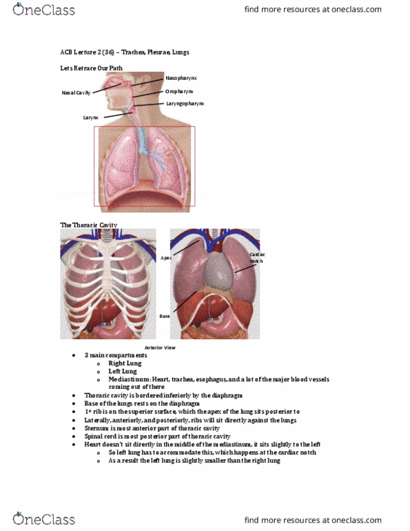
The Thoracic Cavity
●Left lung
○ Has to accommodate for the heart (cardiac notch
●Right lung
●Mediastinum
○ Middle of the thoracic cavity
○ Heart, trachea, esophagus, major blood vessel
●Diaphragm
○ Inferior border
○ Base of the lungs rest on the diaphragm
●1st rib
○ Superior border
○ Apex of the lung
●Sternum
○ Most anterior area
●Spinal cord
○ Most posterior area
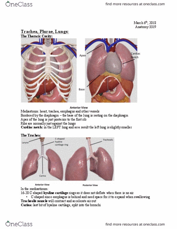
The Trachea
●Anterior View
○ C-shaped hyaline
cartilage rings
■ Protects
and inflates the trachea
■ Not a full circle because it leaves room for the esophagus to expand
when swallowing food
●Posterior View
○ Trachealis
■ Smooth muscle
■ Accelerated the air when you sneeze
■ Sits on the posterior side
●Carina
○ Last little bit of hyaline cartilage, before branching into bronchioles
find more resources at oneclass.com
find more resources at oneclass.com

Internal Structures of the Trachea
● Trachealis
● C-shaped hyaline cartilage ring
● Goblet cells
○ Secrete mucus
■ Traps pathogens and particles
● Respiratory epithelium
○ Has cilia
■ Beats to transport mucus, propels the mucus up and out of the respiratory
tract
■Mucociliary escalator
Tracheostomy
● If done improperly leads to:
○ Rupture of the cardiac
arteries and veins
○ Cutting of the nerves
that innervate the vocal
cords (loss of speech)
● Cut a small incision in the
tracheal rings, insert a
tracheostomy into the treach
○ Allows air bypass in an
obstructed trachea
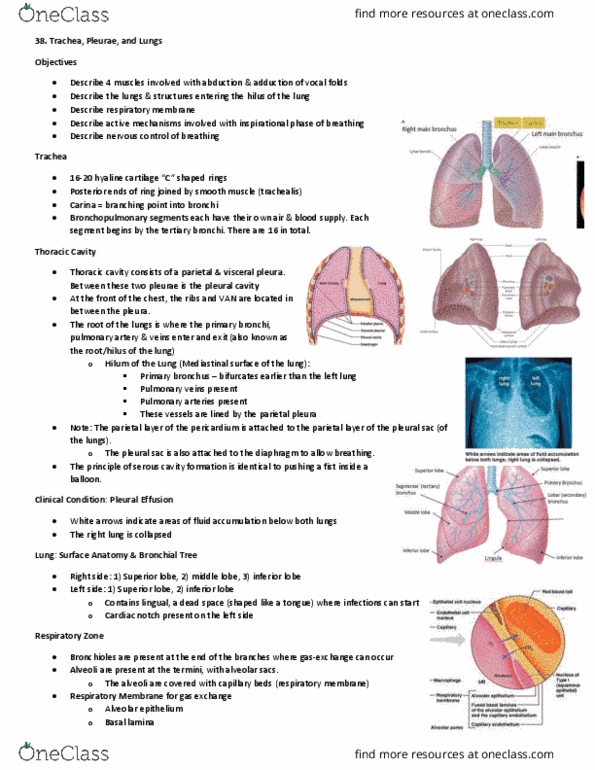
Primary Bronchi
● Branching of the trachea into
find more resources at oneclass.com
find more resources at oneclass.com

the primary occurs around the sternum angle
● Runs through the mediastinum, enter the medial aspect of the lungs
● Objects are more likely to get stuck in the right bronchus
○ Because it is large and
on a more vertical
angle
● Still have regular cartilage and
respiratory epithelium that
makes mucus
○ These features
disappear later
Hilum
● Indentation of the lungs where
all the vessels, lymphatics,
and nerves enter the lung
○ “Root” of the lung
Secondary/lobar Bronchi
● Right lung
○ 3 branches (for 3 lobes)
■ Each secondary
bronchus supplies a
lobe of the lung
■ Superior lobar
bronchus, middle
lobar bronchus,
inferior lobar bronchus
○ 2 fissures
■ Separates the lobes of
the lung
■ Horizontal fissure
● Separates the
superior and middle lobe
find more resources at oneclass.com
find more resources at oneclass.com
Document Summary
Describe the passageway of air through the respiratory system. Identify and describe the anatomical features of the respiratory system and their relationships. Describe the respiratory membrane and the mechanism of gas exchange. Describe how the nervous system regulates breathing. Has to accommodate for the heart (cardiac notch. Base of the lungs rest on the diaphragm. Not a full circle because it leaves room for the esophagus to expand when swallowing food. Last little bit of hyaline cartilage, before branching into bronchioles. Beats to transport mucus, propels the mucus up and out of the respiratory tract. Rupture of the cardiac arteries and veins. Cutting of the nerves that innervate the vocal cords (loss of speech) Cut a small incision in the tracheal rings, insert a tracheostomy into the treach. Allows air bypass in an obstructed trachea. Branching of the trachea into the primary occurs around the sternum angle. Runs through the mediastinum, enter the medial aspect of the lungs.