Anatomy and Cell Biology 3319 Lecture Notes - Lecture 32: Circulatory System, Aorta, Pulmonary Circulation

6 May 2019
School
Department
Professor
Document Summary
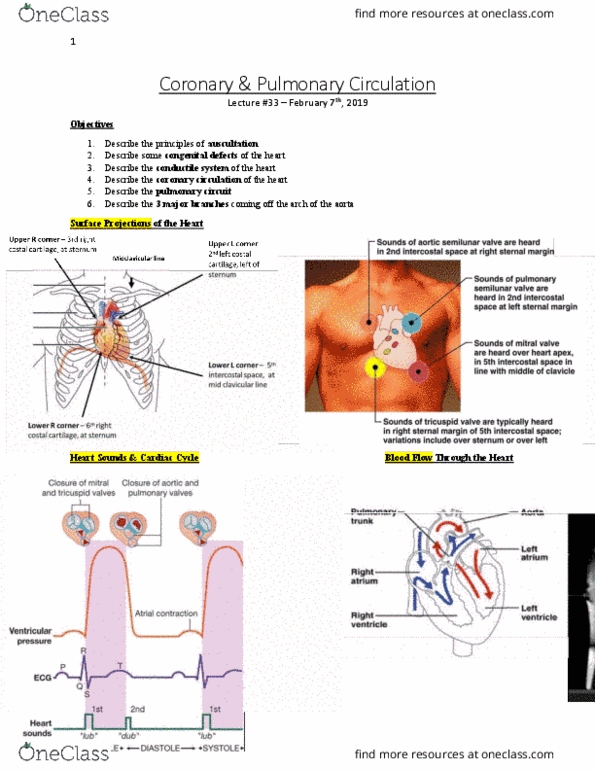
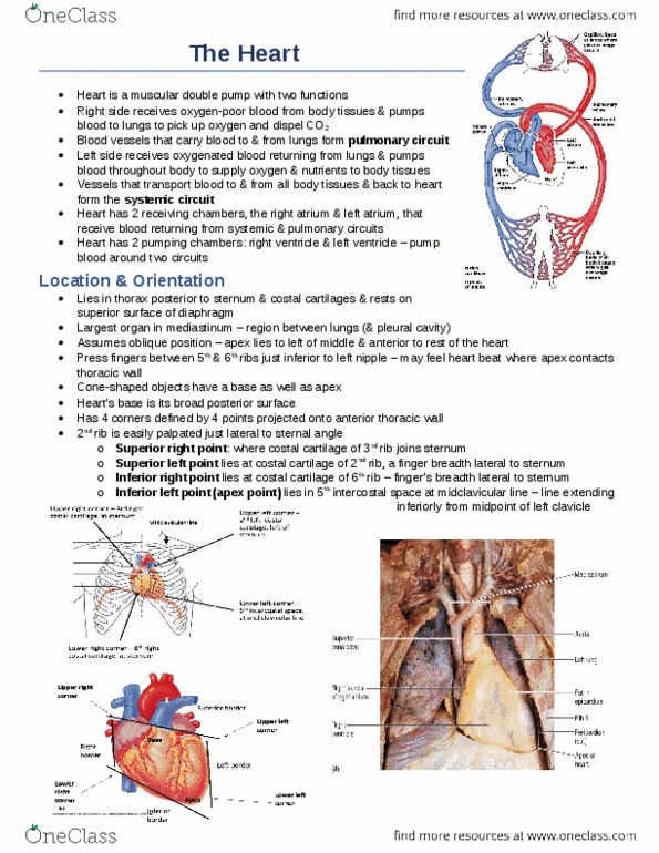
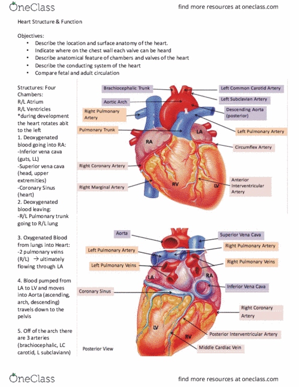
Lecture #32 february 5th, 2019: describe the location and surface anatomy of the heart, describe anatomical features of chambers and valves of the heart, describe the conducting system of the heart, compare fetal and adult circulation. Indicate where on the chest wall each valve can be heard. Consists of: 4 borders, 4 corners, a base, and an apex. The heart is located within a pericardial sac in the middle mediastinum. Pericardial sac consists of: visceral + parietal layers: parietal layer has 2 components, outer fibrous. Fibrous pericardium attaches the pericardial sac to the: great vessels, pleural of lungs, diaphragm. The pericardial cavity lies btwn the visceral & parietal layers of serous pericardium. Analogous to hand pushing into a balloon: fist covered by layer of rubber, pushes further to become enclosed w/in a 2-layered cavity. Heart (fist) pushes into serous body cavity (balloon) from behind: becomes enclosed w/in a 2-layered cavity (pericardial cavity)