Kinesiology 2222A/B Lecture Notes - Lecture 6: Conus Medullaris, Coccyx, Intervertebral Foramina

27 Sep 2018
School
Department
Course
Professor
Document Summary
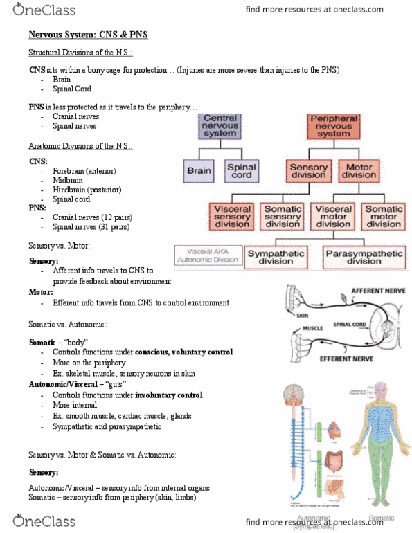
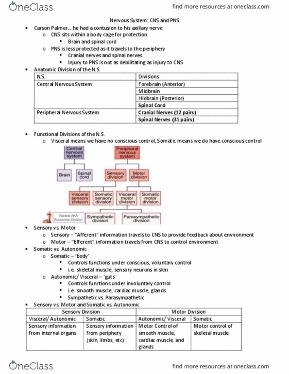
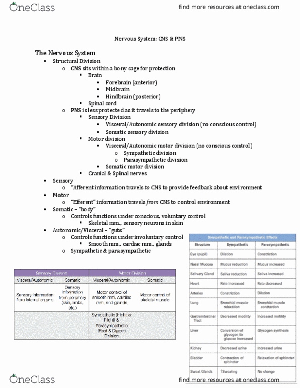
Sits within a body cage for protection. Brain: forebrain (anterior, midbrain, hindbrain (posterior) Info travels to cns to provide feedback about environment. Less protected as it travels to the periphery. Conversion of glycogen to glucose increased glycogen synthesis. Discuss main anatomical features of the spinal cord. Similar organization of cells, but slightly different. Grey matter: nerve cell bodies (neurons) and non-myelinated nerve fibers, grey matter is outside of brain but middle of spinal cord. White matter: myelinated nerve fibers: white matter is inside of brain but outside of spinal cord, myelinate and insulates nerves for better conduction. Extend from foramen magnum to l2 vertebrae. Conus medullaris: cone shaped termination of spinal cord. Cauda equina: bundle of spinal roots become l2 vertebrae: hang off of conus medullaris. Cross-section: h-shaped grey matter surrounded by white matter. Cervical and lumbar enlargement: due to nerves controlling upper and lower limbs.