FORS 3331 Lecture Notes - Lecture 9: Mandibular Canal, Masseter Muscle, Temporal Muscle

1 May 2017
School
Department
Course
Professor
Document Summary
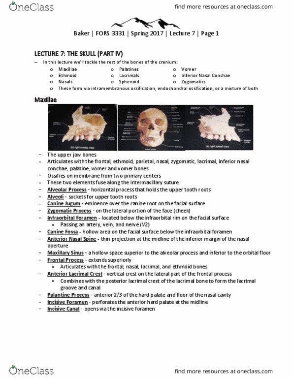
Baker | fors 3331 | spring 2017 | lecture 9 | page 1. Holds lower teeth and has attachment areas for chewing muscles. Articulates with the temporal at the temporomandibular joint. Ossifies as two separate bones: fuse along the mental symphysis in the first year. At birth, the mandible holds un-erupted deciduous teeth. Body - the part that holds the lower teeth: hard, dense, and resistant to destruction, corpus, horizontal ramus. Mental foramen - large hole on the lateral portion of the corpus: below the premolars, transmits the inferior alveolar nerve, part of the mandibular nerve, v3. Mylohyoid line - attachment crest for the mylohyoid muscle: crosses the medial corpus obliquely, starts near the last molar and runs anteriorly and inferiorly. Submandibular fossa - hollow on the medial corpus. Inferior to the mylohyoid line: channel for the submandibular gland (salivary)