SAR HS 369 Lecture Notes - Lecture 10: Abdominopelvic Cavity, Abdominal Cavity, Thoracic Cavity

2 Jul 2020
School
Department
Course
Professor
Document Summary
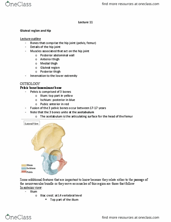
Part of a larger cavity known as the abdominopelvic cavity. The location of most digestive organs, part of the urogenital system (kidneys and most of the uterers) and the spleen. Separated from the thoracic cavity by the thoracic diaphragm. Under cover of the thoracic cage superiorly. Enclosed anterolaterally by multi-layered, musculoaponeurotic, abdominal walls. The three flat muscles of the anterolateral abdominal wall are the: external oblique, internal oblique, transverse abdominis. All three flat muscles end anteriorly in a strong, sheet-like aponeurosis. The aponeuroses of these muscles interlace at the linea alba (la) highlited by the yellow arrow in the image blow. For a broad muscle, like these three ones, rather than the muscle fibers condensing down in a cord-like tendon, the muscle fibers transition to a flat sheet of tissue known as the aponeurosis. The pelvis serves as the site of attachment for some of the muscles of the anterior abdominal wall.