LIFE 102 Lecture Notes - X-Ray Crystallography, Dna Replication, Radiography

Document Summary
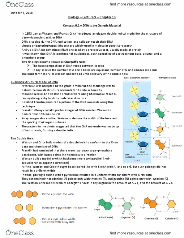
Building a structural model of dna: scientific inquiry. After most biologists became convinced that dna was the genetic material, the challenge was to determine how its structure accounts for its role. Maurice wilkins and rosalind franklin were using a technique called x-ray crystallography to study molecular structure. Franklin produced a picture of the dna molecule using this technique. Franklin"s x-ray crystallographic images of dna enabled watson to deduce that dna was helical. The x-ray images also enabled watson to deduce the width of the helix and the spacing of the nitrogenous base. The width suggested that the dna molecule was made up of two strands, forming a double helix. Watson and crick built models of a double helix to conform to the x-rays and chemistry of dna. Franklin had concluded that there were two antiparallel sugar-phosphate backbones, with the nitrogenous bases paired in the molecule"s interior.