LIFE 210 Lecture Notes - Apaf1, Mitochondrion, Cytoskeleton

Document Summary
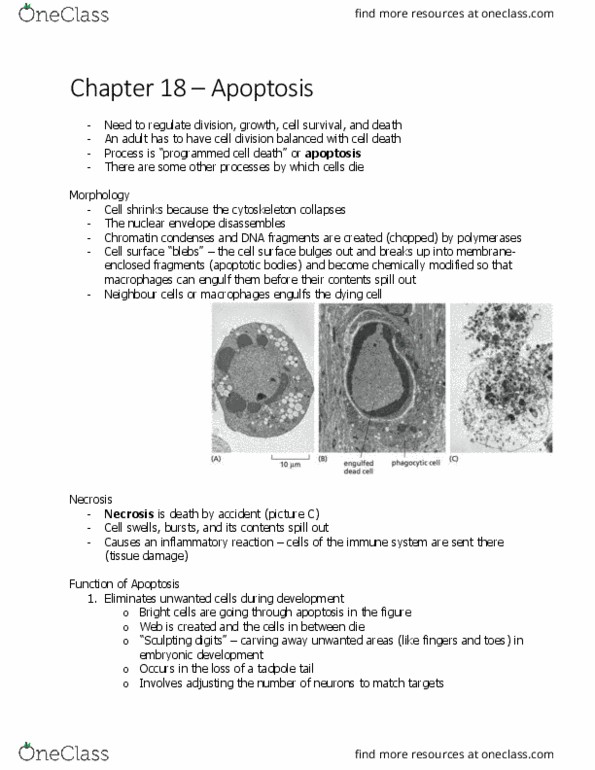
Cells dying by apoptosis undergo characteristic morphological changes. Cells shrink and condense, cytoskeleton collapses, nuclear envelope disassembly, chromatin condenses and fragments. Necrosis: cells die by acute insult, they swell and burst. Half of the neurons normally die soon after they are formed. Example 1: sculpting the digits in a developing mouse paw. Example 2: metamorphosis of a tadpole into a frog. Quality control: eliminate cells that are abnormal, potentially dangerous (some lymphocytes), nonfunctional, damaged (dna damage) Phosphatidylserine flips from inner to outer leaflet of plasma membrane. Release of mitochondria proteins from the intermembrane space. Apoptosis depends on an intracellular protelytic cascade: caspases. Targets: nuclear lamin, cytoskeletal proteins, cell-cell adhesions proteins. Cell surface death receptors activate the extrinsic pathway of apoptosis. Release of cytochrome c from mitochondria during apoptosis. Response to injury or stress such as dna damage, lack of oxygen or nutrients. Cytochrome c binds to apoptotic protease activating factor-1 (apaf1)