BIOG 1440 Lecture Notes - Lecture 17: Endoplasmic Reticulum, T-Tubule, Skeletal Muscle

27 Apr 2018
School
Department
Course
Professor

Focus: Muscles (Skeletal and Smooth)
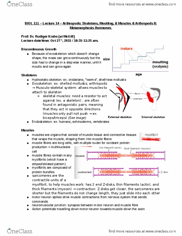
Three Types of Skeleton
Type
What is it?
Examples
Hydrostatic
Body cavity is kept under pressure by
antagonistic circular and longitudinal
muscles
Worms, insect larvae, some
mollusks (cephalopods)
External
(exoskeleton)
Cuticle is generally hard, but is a flexible
membrane at joints. Antagonistic muscles
span joints and attach to stiff cuticle
Arthropods, most molluscs,
brachiopods
Internal
(Endoskeleton)
Flexible joints are held together by
proteinaceous ligaments. Antagonistic
muscles span the joint and attach to stiff
plates or bones
Deuterostomes (echinoderms
and chordates)
Skeletal Muscle--Anatomy
1. Vertebrate skeletal muscle moves bones and body
a. Characterized by hierarchy of smaller and smaller units
2. Skeletal muscle=bundle of long fibers, each a single cell, running parallel to length of
muscle
a. Multinucleate, lots of mitochondria
b. Each muscle fiber is a bundle of smaller myofibrils arranged longitudinally
3. Structure of a skeletal muscle
a. Muscles receive about 20% of cardiac output at rest and this increases with
exercise
b. Single cells are surrounded by endomysium
c. Fascicle: group of muscle cells surrounded by perimysium
i. Whole muscle surrounded by epimysium
d. Blood vessels are located between fascicles but are located inside perimysium
4. The inside of a skeletal muscle cell
a. Sarcolemma is the cell membrane but the endomysium is a layer of CT that
wraps around fiber
b. Single cells are packed w/ myofibrils which are not present in other cell types
c. Many mitochondria distributed throughout length of fiber receiving oxygen
released from hemoglobin in the blood
5. Sarcoplasmic Reticulum (SR): specialized network in muscle cells
a. Surrounds each myofibril
b. Transverse (T)-tubules span from outside down into muscle and associates w/
SR
i. In close contact with region of SR known as terminal cisternae
ii. These regions are the source of much of the calcium stored in SR.
iii. Invaginations of sarcolemma into myofiber
find more resources at oneclass.com
find more resources at oneclass.com
Document Summary
Body cavity is kept under pressure by antagonistic circular and longitudinal muscles. Cuticle is generally hard, but is a flexible membrane at joints. Antagonistic muscles span joints and attach to stiff cuticle. Flexible joints are held together by proteinaceous ligaments. Antagonistic muscles span the joint and attach to stiff plates or bones. In close contact with region of sr known as terminal cisternae. These regions are the source of much of the calcium stored in sr. Skeletal muscle contraction: during contraction, sarcomere length shortens, distance between z-discs decreases during contraction, length of filaments (actin and myosin) do not change but degree of overlap does, myosin is anchored to z-disc by protein called titin. Actin, myosin, titin are most abundant proteins in body. Tropomyosin: proteins that bind end to end to make continuous polymer along actin filament. Stimulus leading to contraction of muscle fiber is an action potential in a motor neuron that makes a synapse with the muscle fiber.