BIOL 22000 Lecture Notes - Lecture 6: Lysozyme, Partial Pressure, Vasodilation

8 Jun 2018
School
Department
Course
Professor

1
Chapter 6 The Respiratory System
6.1 Anatomy and Mechanism of Breathing
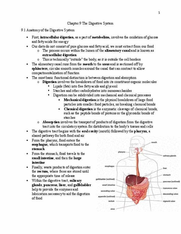
• The lungs are in the thoracic cavity
ANATOMY
• Air enters the respiratory tract through the
external nares of the nose and then passes
through the nasal cavity, where it is filtered
through mucous membranes and nasal
hairs (vibrissae)
• Next, air passes into the pharynx and the
larynx
o The pharynx resides behind the
nasal cavity and at the back of the
mouth
▪ Common pathway for both
air destined for the lungs
and food destined for the
esophagus
o Larynx lies below the pharynx and
is only a pathway for air
▪ To keep food out of the respiratory tract, the opening of the larynx
(glottis) is covered by the epiglottis during swallowing
▪ The larynx contains two vocal cords that are maneuvered using skeletal
muscle and cartilage
• From the larynx, air passes into the cartilaginous trachea and then into one of the two
mainstem bronchi
o The bronchi and trachea contain ciliated epithelial cells to catch material that has
made it past the mucous membranes in the nose and mouth
• In the lungs, the bronchi continue to divide into smaller structures known as bronchioles,
which divide further until they end in the tiny balloon-like structures in which gas
exchange occurs (alveoli)
o Each alveolus is coated with surfactant, a detergent that lowers surface tension
and prevents the alveolus from collapsing on itself
• A network of capillaries surrounds each alveolus to carry oxygen and carbon dioxide
o Capillaries covers the tiny alveolus
• The branching and minute size of the alveoli allow for an exceptionally large surface area
for gas exchange
find more resources at oneclass.com
find more resources at oneclass.com

2
• The lungs are contained in the thoracic cavity, also where
the heart is
• The chest wall forms the outside of the thoracic cavity
• Membranes known as pleurae surround each lung
o Enclose lung as it expands
• The surface adjacent to the lung is the visceral pleura, and
the outer part is the parietal pleura
• The lungs do not fill passively and require skeletal muscle to
generate the negative pressure for expansion
• Diaphragm, a thin, muscular structure that divides the
thoracic (chest) cavity from the abdominal cavity
o The diaphragm is under somatic control
BREATHING
• The space within the sac (between visceral pleura and parietal pleura) is referred to as the
intrapleural space, which contains a thin layer of fluid
o This pleural fluid helps lubricate the two pleural surfaces
• The pressure differentials that can be created across the pleura ultimately drive breathing
Inhalation
• Inhalation is an active process
• We use our diaphragm as well as the
external intercostal muscles (one of
the layers of muscles between the ribs)
to expand the thoracic cavity
• As the diaphragm flattens and the
chest wall expands outward, the
intrathoracic volume (the volume of
the chest cavity) increases
o Since the intrapleural space
closed, as volume increase the
pressure decrease
• The pressure in the lungs is now greater than intrapleural space
• The lung will therefore expand into the intrapleural space, and the pressure in the lung
will degree
• Air will then be sucked in from a higher-pressure environment – the outside world
• This mechanism is referred to as negative-pressure breathing because the driving force
is the lower (relatively negative) pressure in the intrapleural space compared with the
lungs
find more resources at oneclass.com
find more resources at oneclass.com