BIOL 22000 Lecture Notes - Lecture 11: Sodium Channel, Sarcomere, Cisterna

26 Jun 2018
School
Department
Course
Professor

Lecture 11
Outline – excitation-contraction coupling
oAnatomy of Skeletal Muscle
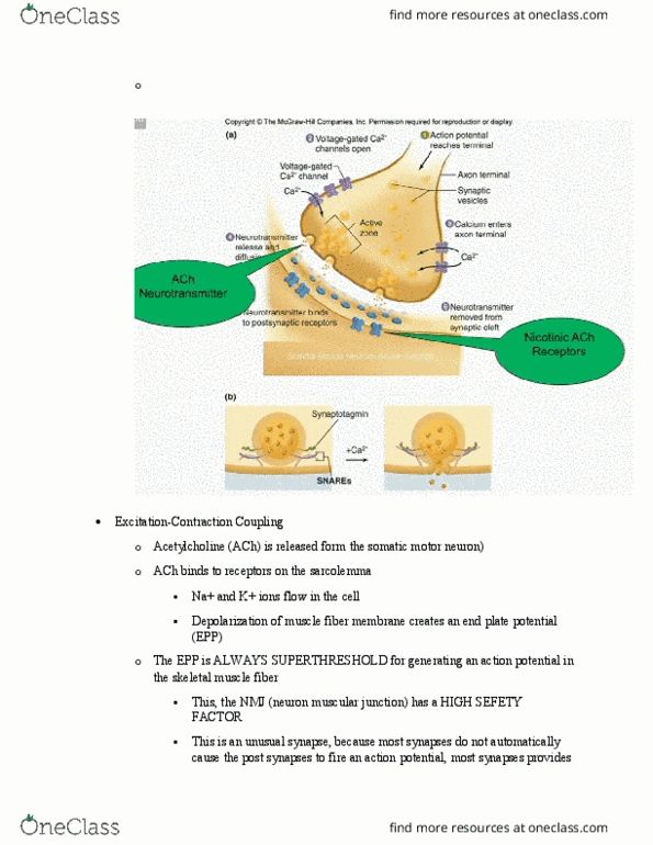
oExcitation – synaptic transmission at the neuromuscular junction
oCoupling – release of intracellular calcium
oContraction – sliding filament theory
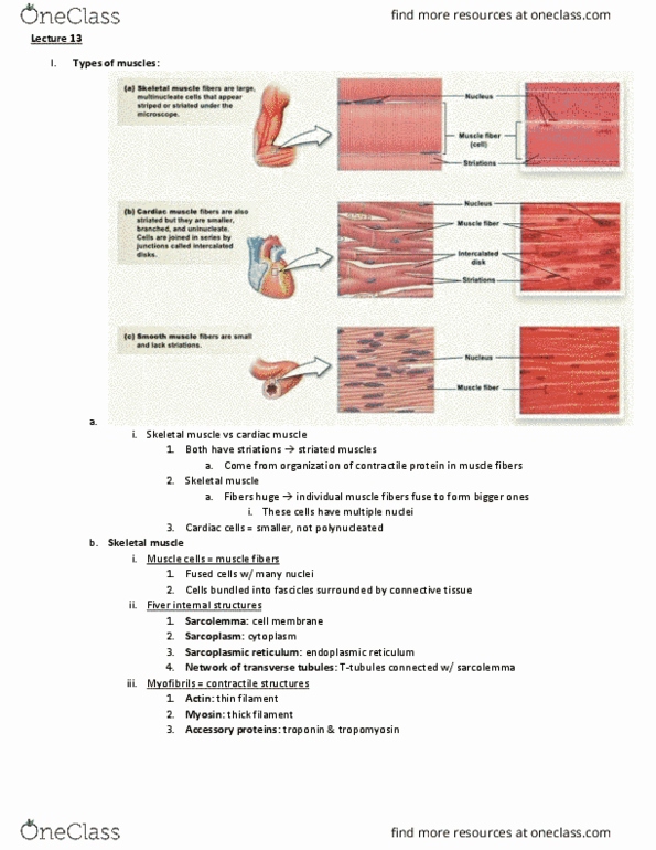
oBoth skeletal muscle and cardiac muscles are striated muscle
oA unique characteristic is that skeletal muscles is that fiber cell is large
oCardiac cells are smaller and are not polynucleated
Anatomy of Skeletal Muscle
oMuscle cells are called muscle fibers
Fused cells with many nuclei
find more resources at oneclass.com
find more resources at oneclass.com

Cells bundled into fascicles sorrunded by connective tissue
oFiber internal structures
Sarcolemma: cell membrane
Sarcoplasm: cytoplasm
Sarcoplasmic reticulum: endoplasmic reticulum
Network of transverse tubules: T-tubules connected with the sarcolemma
This allows plasma membrane to go all the way through the inside
of the muscle cells
Because the plasma membrane can generate action potential, the
action potential can conduct all the way to the inside of the muscle
cell, and bring the excitation all the way in the center of the muscle
fiber
oMyofibrils are the contractile structures
Actin: thin filament
Myosin: thick filament
Accessory proteins: troponin and tropomyosin
find more resources at oneclass.com
find more resources at oneclass.com

oThe nerve is the axon that is coming down to control the muscle fiber
Structure of a Skeletal muscle Fiber
find more resources at oneclass.com
find more resources at oneclass.com
Document Summary
Anatomy of skeletal muscle: muscle cells are called muscle fibers. Cells bundled into fascicles sorrunded by connective tissue: fiber internal structures. Network of transverse tubules: t-tubules connected with the sarcolemma. This allows plasma membrane to go all the way through the inside of the muscle cells. Accessory proteins: troponin and tropomyosin: the nerve is the axon that is coming down to control the muscle fiber. T-tubules are just sarcolemma embedded in the muscle cells. When there is an action potential, it propagates down the t-tubule and go all the way inside. Sarcoplasmic reticulum is the calcium storage in muscle cells. When muscle cells are excited, sarcoplasmic reticulum release calcium right on top of myofibril. Myosin head bind atp, so they are atpase. Myosin head has binding site for actin: actin has two forms. G-actin molecule each individual represent globule actin, also bind site.