PSYCH 212 Lecture Notes - Lecture 3: Multiple Sclerosis, Neuroglia, Optogenetics

11 Jan 2017
School
Department
Course
Professor
Document Summary
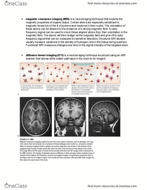
Structural neuroimaging structural magnetic resonance imaging (mri) - generates static high resolution images of tissue. Indexes distribution of hydrogen proteins because hydrogen covaries with tissue density diffusion tensor imaging (dti) - mri technique that generates imaged of the pathways or white matter tracts in the brain. Indexing the myelin sheath which wraps around the axon. Generates images by assessing the mobility of water molecules along white matter tracts. Important to multiple sclerosis because it"s when you start losing the myelin. A limitation of structural neuroimaging is that it generates static image of brain tissue. In order to asses brain function researchers use functional neuroimaging techniques. When nerve cells are activated they increase the consumption of energy which results in increased blood ow. Fmri indexed this distribution of blood ow to make inferences about the level of activation of neural tissue.