BIOL 3312 Lecture Notes - Lecture 2: Transmission Electron Microscopy, Scanning Electron Microscope, Platinum Film

Document Summary
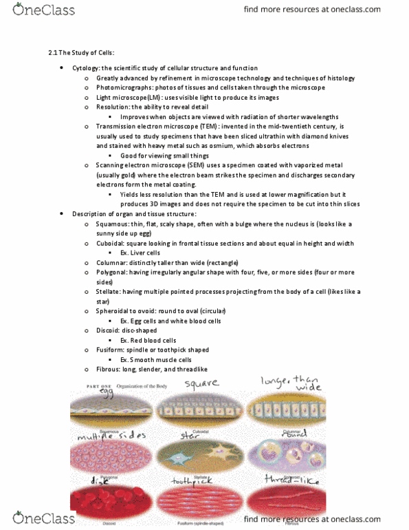
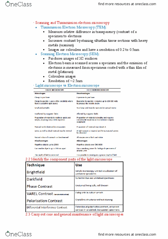
Cytology is the scientific study of cellular structure and function. It is important so we know how the cell works because our body is made up of cells. Light microscope: easiest to use, least expensive, most often used, may only magnify up to. It uses visible light to produce its images. most limited for magnification. Transmission electron microscope: used to view specimen that have been sliced ultra thin and stained with a heavy metal. Good enough to see things as small as proteins, nucleic acids, and other large molecules. two-dimensional black-and-white images. Scanning electron microscope: uses a specimen coated with vaporized metal (gold). Electron beam hits the specimen and discharges secondary electrons from the metal coating. The electrons hit a fluorescent screen and produce an image. only see surface of specimens. Freeze-fracture a specimen is frozen rapidly and cracked on a plane through the tissue. After cleaving, both surfaces are shadowed with a platinum film.