01:119:116 Lecture Notes - Lecture 15: Pear-Shaped, Myometrium, Oogenesis

17 Aug 2016
School
Department
Course
Professor
Document Summary
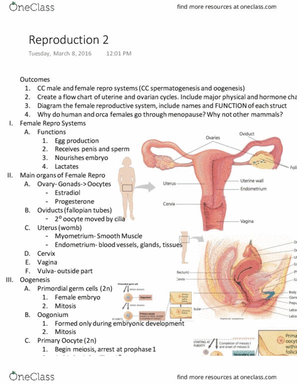
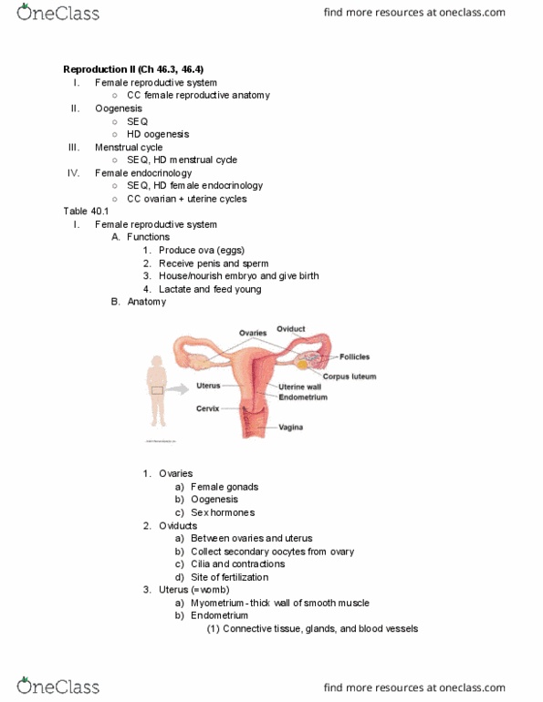
I. outcomes: by the end of reproduction, you should be able to compare and contrast the male and female reproductive, as well as cc spermatogenesis and oogenesis, create a flow chart of the uterine and ovarian cycles. A. functions: produces ova (eggs), receives penis and sperm, houses and nourishes developing embryo; give birth, lactates and feeds young. Iii. main organs of female reproduction (important: figure 46. 10) A. ovary- gonads -> oocytes, estrodiol and progesterone: ovaries- female gonad, located in abdominal cavity, held in place by several ligaments, produce, gametes via oogenesis, sex hormones- estradiol and progesterone. D. cervix: cervix- lower part of the uterus, closes uterus and separates from vagina, common site of cancer, >90% cases of cancer due to hpv. E. vagina (birth canal: vagina- elastic muscular tube, extends to outside of body, receives penis and sperm, birth canal. F. vulva: vulva- outside part that surrounds the opening of the vagina, external genitalia, covers external openings. A. primordial germ cells (2n: female embryo, mitosis.