01:146:356 Lecture Notes - Lecture 11: Hemoglobin, Action Potential, Preganglionic Nerve Fibers

23 Mar 2016
School
Department
Course
Professor
Document Summary
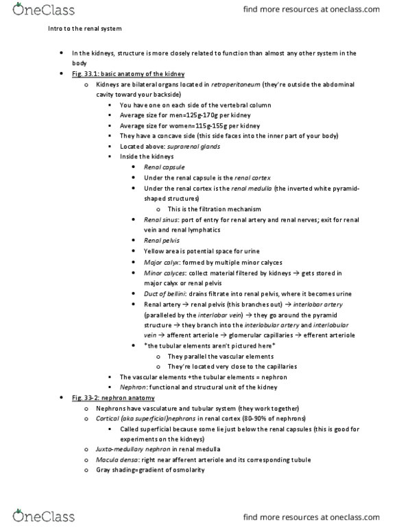
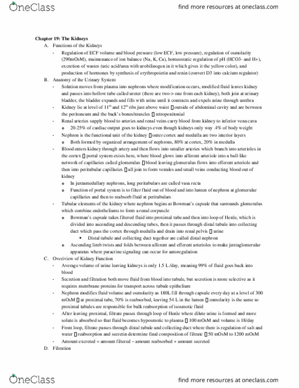
Functional anatomy and basic elements elements of renal physiology. Location, structure and four basic functions of kidneys. Blood supply to kidneys and superficial vs juxtamedullary nephrons. Structure of the mammalian nephron (illustrating papillary zones and the location of glomerular capillaries) fig. Both kidneys combined is less than 1/10th of body mass 125-126g each kidney. Inner epithelial lining of the abdominal cavity: peritinium--> kidneys lie behind them. Start longitudinal- 12 vertebrae, extend down to the 3rd/4th lung vertebrae. Kidney is a bean shaped structure, convex and concave surfaces. On the concave surface there"s an indentation that we call the renal hylis- port of entry for renal artery and renal nerves and it"s the port of exit for the renal vein and lymphatic vessels. Because of the structure of these combined elements, they create an indentation. - 4 layers of tissue- outer fibris connective tissue (renal capsule) And just beneath the renal capsule is the renal cortex, below is renal medulla.