BSC 215 Lecture Notes - Lecture 33: Round Window, Ear Canal, Ossicles

11 Jun 2018
School
Department
Course
Professor

Hearing
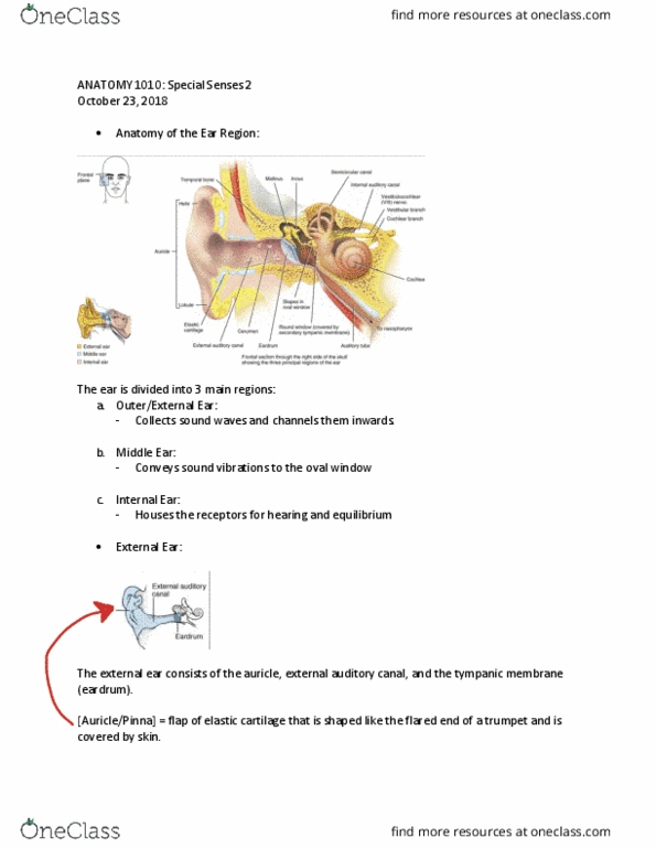
The organ of hearing, the ear, consists of three major regions, shown in
Figure 1.
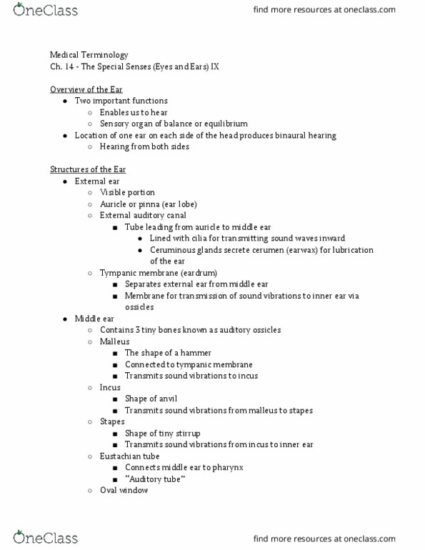
• The outer (external) ear consists of the auricle (pinna), a flap of elastic
cartilage that protrudes from the head, and the external auditory canal
(meatus), a tube that enters the temporal bone. The canal is lined with
ceruminous glands that secrete cerumen (earwax), a sticky substance
that traps dirt and other foreign objects. The eardrum (tympanic
membrane), at the internal end of the external auditory canal, vibrates
in response to incident sound waves.
• The middle ear (tympanic cavity) is an air‐filled cavity within the
temporal bone. It contains three small bones, the auditory ossicles.
These bones, called the malleus, incus, and stapes, act as a lever
system that amplifies and transfers vibrations of the eardrum to the
inner ear. The malleus at one end connects to the eardrum, while the
stapes at the other end attaches with ligaments to the oval window, a
small, membrane‐covered opening into the inner ear. Synovial joints
connect the incus, the center bone of the auditory ossicles, to the
malleus and stapes on each side. A second membrane‐covered
opening to the inner ear, the round window (secondary tympanic
membrane), lies just below the oval window. A third opening leads to
the auditory (Eustachian) tube, which connects the middle ear to the
upper throat. The auditory tube allows pressure differences between
the middle and outer ear to equalize, thus reducing tension on the
eardrum. Two muscles in the middle ear, the tensor tympani and the
stapedius, connect to the malleus and stapes, respectively. Contraction
of these two muscles restricts the movement of the eardrum and
auditory ossicles, reducing damage that may occur when they are
exposed to excessive vibration from loud noises.
• The inner (internal) ear, also called the labyrinth, is a system of
double‐walled canals. The canals consist of an outer bony (osseous)
labyrinth that encloses an inner membranous labyrinth. Perilymph fills
the space between the two labyrinths, and endolymph fills the inner
labyrinth. This double‐layer labyrinth structure is found throughout the
following inner ear structures. This labyrinth is made of three
semicircular canals and a snail‐shaped cochlea (see Figure 1).
find more resources at oneclass.com
find more resources at oneclass.com

Figure 1. The three major regions of the ear are the outer ear, the middle
ear, and the inner ear.
find more resources at oneclass.com
find more resources at oneclass.com
Document Summary
The organ of hearing, the ear, consists of three major regions, shown in. Figure 1: the outer (external) ear consists of the auricle (pinna), a flap of elastic cartilage that protrudes from the head, and the external auditory canal (meatus), a tube that enters the temporal bone. The canal is lined with ceruminous glands that secrete cerumen (earwax), a sticky substance that traps dirt and other foreign objects. The eardrum (tympanic membrane), at the internal end of the external auditory canal, vibrates in response to incident sound waves. temporal bone. It contains three small bones, the auditory ossicles. These bones, called the malleus, incus, and stapes, act as a lever system that amplifies and transfers vibrations of the eardrum to the inner ear. Synovial joints malleus and stapes on each side. A second membrane covered opening to the inner ear, the round window (secondary tympanic membrane), lies just below the oval window.