BSCI 330 Lecture Notes - Lecture 2: Fluorophore, Scanning Electron Microscope, Total Internal Reflection Fluorescence Microscope

2 Sep 2016
School
Department
Course
Professor
Document Summary
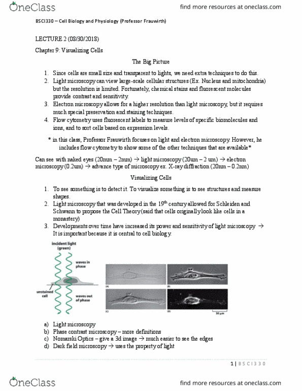
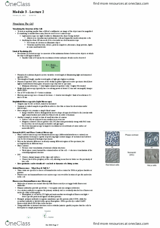
Bsci330 lecture 2 visualizing cells (ch. 1st weekly quiz will be available tomorrow (due monday 9/5 at midnight) There will be 30 minutes to complete the quiz. In order to understand cellular function requires techniques for visualizing individual cells: cells are small, cells are usually transparent to life so they"re difficult to see with our own eyes; we want to have tools to see cells. Light microscopy is used to image large-scale cellular structure: disadvantage: resolution is limited, uses chemical stains and fluorescent molecules for contract and sensitivity. Electron microscopy allows much higher resolution than light microscopy: disadvantage: large and expensive, requires special preservation and staining techniques. Cellular structures are too small to see with the naked eye. Cellular structures need to be magnified in order to study them. The bulk of cell biology is being able to see cellular structures and having the ability to visualize cells. Modifications to light microscopy are still ongoing.