ATH 1061 Lecture Notes - Lecture 21: Anterior Cruciate Ligament Injury, Intercondylar Area, Medial Condyle Of Tibia


Knee Injuries
Chapter 20
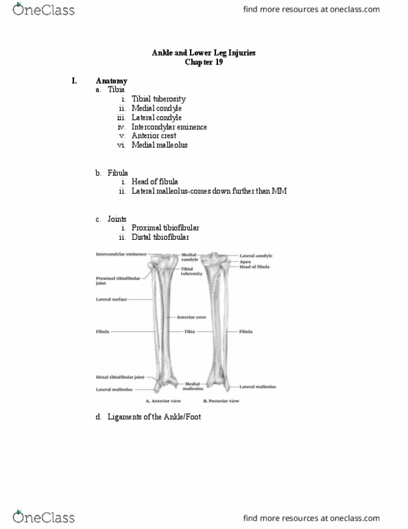
I. Knee Bony Anatomy
a. Femur
i. Medial epicondyle
ii. Lateral epicondyle
iii. Medial condyle
iv. Lateral condyle
v. Intercondylar fossa
b. Tibia
i. Medial condyle
ii. Lateral condyle
iii. Intercondylar eminence
iv. Gerdy’s tubercle
v. Tibial tuberosity
c. Fibula
i. Head of fibula
ii. Apex
d. Patella
i. Articular facets
ii. Base
iii. Apex
II. Knee Structural Anatomy
a. Anterior Cruciate Ligament (ACL)
b. Posterior Cruciate Ligament (PCL)
c. Medial Collateral Ligament (MCL)
d. Lateral Collateral Ligament (LCL)
e. Medial Meniscus
f. Lateral Meniscus
g. Transverse Ligament
h. Ligament of Wrisberg
i. Ligament of Humphrey
j. Middle Genicular Artery
find more resources at oneclass.com
find more resources at oneclass.com
Document Summary
Knee bony anatomy: femur, medial epicondyle, lateral epicondyle, medial condyle, lateral condyle, intercondylar fossa, tibia, medial condyle, lateral condyle, intercondylar eminence, gerdy"s tubercle, tibial tuberosity, fibula, head of fibula, apex, patella, articular facets, base, apex. Knee structural anatomy: anterior cruciate ligament (acl, posterior cruciate ligament (pcl, medial collateral ligament (mcl, lateral collateral ligament (lcl, medial meniscus, lateral meniscus, transverse ligament, ligament of wrisberg, ligament of humphrey, middle genicular artery. Knee anatomy: bursa, infrapatellar, suprapatellar, prepatellar, superficial, dynamic stabilizers, quadriceps muscles, vastus lateralis, vastus intermedius, function, location, crosses hip and knee joint, hamstring muscles, rectus femoris, function, location, crosses the hip and knee joint. Knee extensor mechanism: pulley mechanism, q-angle . Knee injuries: acute, patella subluxation or dislocation, etiology, deceleration with simultaneous cutting in opposite direction (valgus force at knee, some athletes may be predisposed to injury, signs and symptoms, with subluxation, pain and swelling, restricted.