BIOL 171 Lecture Notes - Lecture 31: Renal Pyramids, Nephron, Renal Cortex

Document Summary
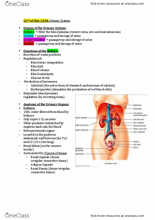
Anatomy of the urinary system: renal anatomy/urinary system (retroperitoneal, renal physiology/urinary, associated lab values, normal varians, sonographic appearance. Renal anatomy / urinary system (retroperitoneal: 2 kidneys, 2 ureters, urinary bladder, urethra, male prostastic, membranous, and spongy/penile urethra, females small urethra, retroperitoneal areas kidney, urethra, ivc, pancreas, 2nd duodenum, etc. Kidneys: right kidney lies slightly lower than the left kidney because the large right lobe of the liver pushes it inferiorly, both kidneys move downward approx. 1 inch: dark red, bean shaped organs, lateral surface is convex surface and medial concave, measurement, 9 to 12cm long, 5cm wide, 2. 5 cm thick, outer cortex darker than the inner medulla bc of increased blood perfusion. Renal sinus is composed of renal hilum, connective tissue, calyces and renal pelvis, The renal fascia (aka gerota fascia) is a layer of connective tissue encapsulating the kidneys. Left adrenal gland: spleen, stomach, and pancreas. Left colic flexure: coils of jejunum, note: liver capsule glisson"s capsule.