SLHS 2203 Lecture Notes - Lecture 4: Effusion, Spirometry, Poliovirus

8 Feb 2018
School
Department
Course
Professor
Document Summary
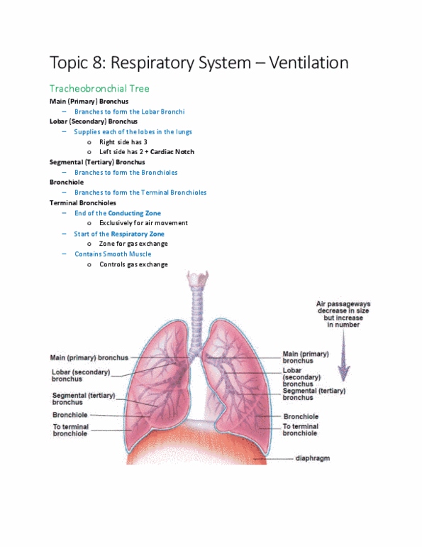
Moving our way down to the lungs. Series of cavities and tubes that conduct air into the lungs. Distal to the terminal bronchioles, the branches of the respiratory tree are now called respiratory bronchioles. These end in alveoli (air sacs) surrounded by capillary beds. A balloon-like sacs surrounded by a bed of capillaries. Left and right lungs are not symmetrical. The left lung has a cardiac notch; this is where the heart sits. The left lung has two lobes, but the right lung has three . Two fluid-filled spaces around each lung (left/right) The fluid-filled space between pleural cavities, contains pericardial cavity. The lungs are double-bagged with a bilayer membrane! Layers are separated by a thin space called the serosa cavity filled with a fluid (called serosa) that functionally couples the two layers so they move together. Examples of pleural disorders: pleural effusion, pleurisy, pneumothorax.