COGS 17 Lecture Notes - Lecture 2: Cranial Nerves, Prefrontal Cortex, Central Canal

16 Nov 2016
School
Department
Course
Professor
Document Summary
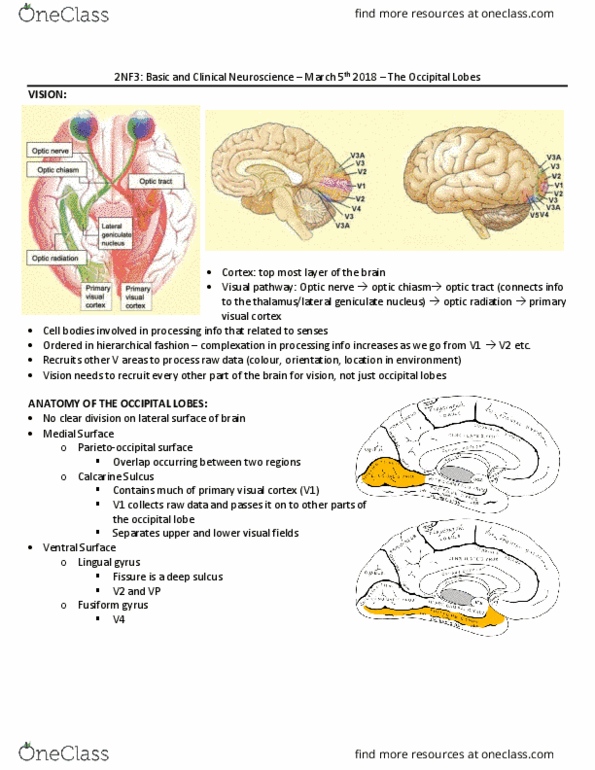
Lecture 1b: anatomy of the nervous system continued. A six-layer sheet of cells, unfolded= < 1meter square. Can use nissl stain to see cell bodies. Info projected to cortex enters at level 4. Central sulcus: major divide from front to back. Lateral fissure: deeper than a sulcus, runs along the side. At rear most part of the brain, has v1 (primary projection area from thalamus) Ventral pathway- who/what , recognizes color and detail. Dorsal pathway- where/how involved with visual info, where things are/ how to engage, motion and depth. Medial temporal (mt) along dorsal pathway, visual pathway to parietal lobe, includes direction sensitive motion detectors. Next is medial superior temporal (mst), includes optic flow detectors, as you move visual field expands/contracts with forward/backward motion. A1 primary projection area for audition from thalamus, concerned with frequency and loudness. Emotional expression and interpretation, this is right hemisphere dominant, anterior part of temporal lobe.