PSYCH 345 Lecture Notes - Lecture 13: Visual System, Parietal Lobe, Temporal Lobe

Document Summary
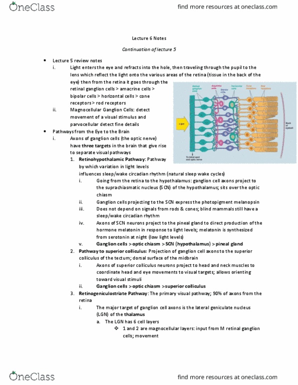
What pathway: ventral path, temporal lobe, parvocellular pathway, retinal p-cells (ganglion cells in the lgn make up the layers, slow conducting, acuity (to figure out what it is, color (fovea dominant, 2 layers for each eye (superior layers, smaller) Where pathway: dorsal, eye v1 parietal lobe, magnocellular pathway, retinal m-cells (ganglion cells in the lgn make up the layers) **also project to superior colliculus: fast conducting, shapes, black and white (peripheral, 1 layer from each eye projecting to inferior layers of lgn (large) Ungerleider & mishkin: object vs landmark discrimination, temporal vs parietal lesions, bilateral, **double dissociation uses two single associations to negate the possibility of an external factor. Sensory receptor (i. e. eyes) thalamus primary cortex secondary (modality) tertiary (multi-modality, integration area?) Blobs/interblobs make up the parvocellular layers of lgn. V1 is the first place in the visual cortex where inputs from each eye are combined. Hierarchial visual receptive field size increases and increases in complexity.