ACB 8120 Lecture Notes - Lecture 74: Dural Venous Sinuses, Superior Sagittal Sinus, Falx Cerebri

12 Apr 2020
School
Department
Course
Professor
Document Summary
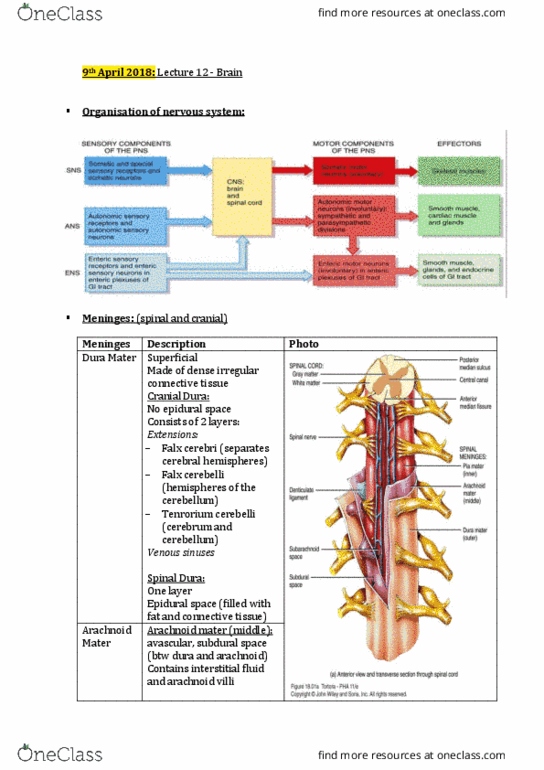
Each of the primary and secondary vesicles retain its original hollow lumen. Pretty much just replace cephalon with cele to get the lumen inside that portion of the neural tube. Outer layer ectomeninx dura mater. Inner layer endomeninx arachnoid and pia mater. Pia = innermost, follows the bumps of the cerebrum. Trabeculae - spider web looking strands between the arachnoid and pia. Villi - cauliflower that projects into the superior sagittal sinus that is the. Shop vac that sucks all the bad stuff out of the subarachnoid space and transports it to the dural venous sinus. Doesn"t follow the grooves of the cerebrum. Periosteal - portion of the dura that is on the bone. Meningeal - portion of the dura that is on the arachnoid and goes down to. Non existent unless injury with fluid accumulation. Between skull and periosteal dura and is non existent unless there is an injury.