ANPS 020 Lecture Notes - Lecture 28: Peritoneal Cavity, Exocrine Gland, Serous Membrane

10 Apr 2019
School
Department
Course
Professor
Document Summary
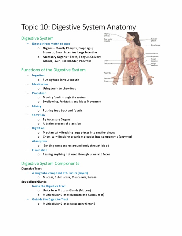
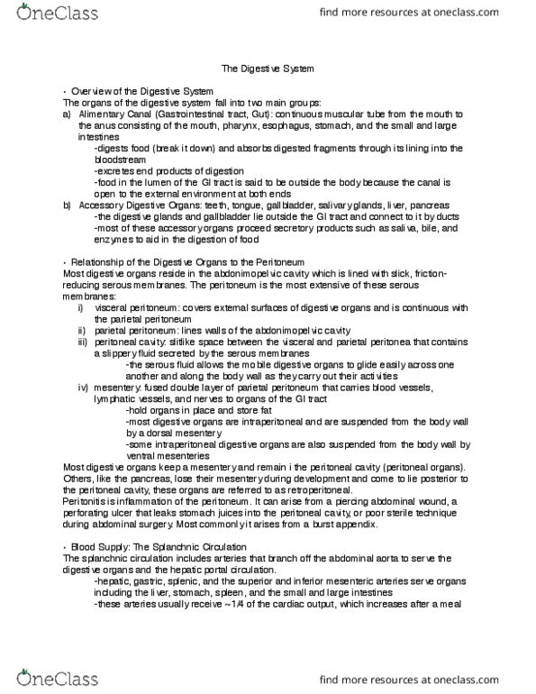
Injestion taking in food and water via the mouth. Populsion movement of food/water by rhythmic contractions of smooth muscle. Digestion mechanically: physically break food into smaller parts. Absorption movement of nutrients from the lumen to blood or lymph. Defacation elimination of solid waste (feces): ingestible substances and metabolic waste. Most digestive organs are intraperitoneal and are suspended from the body wall by a dorsal mesentery. Some intraperitoneal digestive organs are also suspended from the body wall by ventral mesenteries. Some digestive organs are retroperitoneal because they have lost their mesentery during development. Peritoneum serous membrane sac with two layers and cavity. Parietal peritoneum: lines the inner surface of the body wall. Visceral peritoneum: lines the external surface of digestive organs. Peritoneal cavity: fluid-filled potential space between the peritoneal layers. Organs that lie outside the peritoneum are called retroperitoneal. Mesenteries are double layers of peritoneum that extend from the body wall and hold organs in place.