HSE201 Study Guide - Final Guide: Voltage-Dependent Calcium Channel, Neuromuscular Junction, Endoplasmic Reticulum

7 Aug 2018
School
Department
Course
Professor
Document Summary
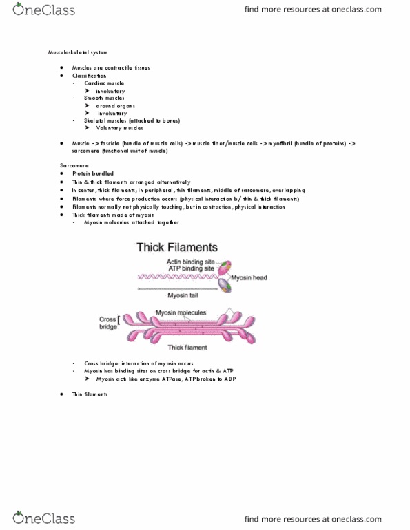
The function and anatomical/structural arrangement of key skeletal muscle components. Muscle fascicle fibre (cell) myo bril sarcomere [contains myosin (thick laments) and actin (thin laments)] Actin: peripherally located in sarcomere, surrounded by regulatory proteins (troponin and tropomyosin) Myosin: centrally located in sarcomere, has a head (binding site for actin) and tail (combines with other tails to form myo lament) segments. Regulatory proteins: at rest myosin binding sites blocked by tropomyosin, when ca2+ present binds to troponin and moves tropomyosin exposing binding sites. Motor unit: a somatic motor neuron and the muscle bres it innervates. Neuromuscular junction: one per cell, synapse b/w motoneuron and muscle bre. Transverse tubules: function to rapidly and evenly distributes action potentials (ap) Sarcoplasmic reticulum: surrounds each myo bril, 2 regions; terminal cisternae (release. Ca2+ into cytosol), longitudinal tubules (uptake ca2+ from cytosol) Fascia (super cial, under skin, covers all body tissues) Sarcolemma (rippled to allow stretching without membrane damage)