HBS2HAB Final: comprehensive-notes-for-hbs2hab


lOMoARcPSD| 1536344
HBS2HAB – HUMAN ANATOMY B
TOPIC 1 AND 2: INTRODUCTION TO UPPER LIMB
MAJOR PRINCIPLES:
E3 Because the upper and lower limbs develop from identical patterns in the embryonic limb bud
the have homologous components
OBJECTIVES:
LO1 – PENTADACTYL LIMB AND THE SKELETAL COMPONENTS OF A HUMAN LIMB
A pentadactyl limb is a limb with 5 digits at the end.
The skeletal components of a typical human limb is as follows:
- Attachment to axial skeleton via girdles
- One bone in proximal segment of limb
- Two bones in distal segment of limb
- Joint complex
- End of limb such as hand/foot with multiple bones
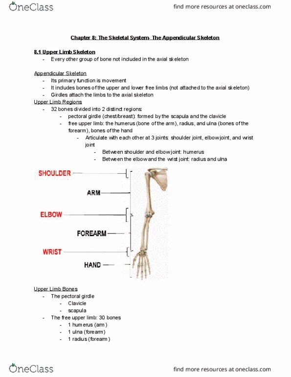
The upper limbs conforms to this pattern as it consists of FOUR major segments:
- Shoulder: Proximal segment of limb that
overlaps with parts of the trunk and lower
lateral neck. It overlies half of the pectoral
girdle.
- Arm: First segment of the free upper limb
(more mobile and independent of trunk) and
longest segment of limb. Extends between
and connects shoulder to elbow and consists
of anterior and posterior regions of arm,
centred around humerus.
- Forearm: Second longest segment of limb
extending between and connecting elbow
and wrist. Also includes anterior and
posterior divisions. Consists of 2 bones –
radius and ulna.
- Hand: Part of upper limb distal to the
forearm that is formed around the carpus,
metacarpus and phalanges. Multiple bones
connecting together to form hand.
1
find more resources at oneclass.com
find more resources at oneclass.com

lOMoARcPSD| 1536344
LO2 – REGIONS OF UPPER LIMB AND HOMOLOGOUS REGIONS IN LOWER LIMB
The pectoral girdle consists of the scapulae and clavicles, connected to the manubrium of the
sternum. The homologous region of the lower limb is the pelvic girdle, consisting of the 2 hip bones
connected to the sacrum.
The arm consists of the humerus, the largest bone in the upper limb, and articulates with the scapula
at the GHJ, and the radius and ulna at the elbow joint. The homologous region of the lower limb is the
femur, the largest bone in the lower limb, articulating with the pelvis and tibia and fibula at the knee
joint.
The hand consists of the carpals, metacarpals and phalanges, connected by a joint complex. The
homologous region of the lower limb is the foot, consisting of the tarsals, metatarsals and phalanges.
The shoulder consists of the pectoral, scapular, and deltoid regions of the upper limb, and the lateral
part of the lateral cervical region, overlying half the pectoral girdle. The homologous region of the
lower limb is the pelvis.
The forearm consists of the radius (shorter bone) and ulna (stabilising bone of forearm) which extend
between the elbow and wrist. The homologous region of the lower limb is the lower leg consisting of
the tibia and fibula, which extend between the knee and ankle joints.
The palm is the central region of the anterior of the hand and consists of the area between the 5
phalanges and the carpus. The homologous region of the lower limb is the sole of the foot.
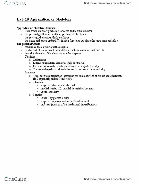
LO3 – UPPER LIMB JOINTS
The sternoclavicular (SC) joint is a saddle type of synovial joint but
functions as a ball and socket joint. It is located between the
clavicle and manubrium of the sternum and is the only attachment
of the upper limb to the axial skeleton. The SC joint allows for a
large degree of mobility and helps with elevation and depression
of the shoulders, protraction and retraction of the shoulders, and
shoulder rotation.
The acromioclavicular (AC) joint is a plane type of synovial joint, located
where the lateral end of the clavicle articulates with the acromion of the
scapula. It allows for a degree of axial rotation and anteroposterior
movement. As no muscles act directly on the joint, all movement is
passive, and is initiated by movement at other joints.
The scapulothoracic joint is a physiological joint, in which movement occurs between musculoskeletal
structures (between scapula and associated muscles and thoracic wall), rather than an anatomical joint,
in which movement occurs between directly articulating skeletal elements. This joint is where the
scapular movements of elevation-depression, protraction-retraction, and rotation occur.
2
find more resources at oneclass.com
find more resources at oneclass.com

lOMoARcPSD| 1536344
The glenohumeral (shoulder) joint (GHJ) is a ball and socket joint
between the scapula and the humerus. It is the major point
connecting the upper limb to the trunk and one of the most mobile
joints in the body. As a ball and socket joint, there is a wide range
of movement permitted such as flexion-extension, abduction-
adduction, and medial-lateral rotation.
The elbow joint oets the proper ar to the forear. Its
marked on the upper limb by the medial and lateral epicondyles,
and the olecranon process. The joint is classed as a synovial hinge
joint. The orientation of the bones forming the elbow joint allows
for extension and flexion of the forearm.
The radioulnar joints are 2 locations in which the radius and ulna
articulate in the forearm. The proximal (superior) radioulnar joint is
located near the elbow, and is an articulation between the head of
the radius, and radial notch of the ulna. The distal (inferior)
radioulnar joint is located near the wrist, and is an articulation
between the ulnar notch of the radius, and the ulnar head. Both of
these joints are classified as pivot joints, responsible for pronation
and supination of the forearm.
The wrist (radiocarpal) joint is a synovial joint marking the
area of transition between the forearm and the hand. It is
formed by the proximal row of the carpal bones (excl.
pisiform) and the distal end of the radius, and the articular
disc. All movements of the wrist are performed by the muscles
of the forearm. These movements include flexion-extension,
and adduction-abduction.
The metacarpophalangeal joints are the condyloid type of synovial
joint that permit movement in 2 planes; flexion-extension and
adduction-abduction. Digits 2-5 allow for flexion-extension,
adduction-abduction, and circumduction movement. Movement of
the thumb is limited to flexion-extension. The interphalangeal joints
are the hinge type of synovial joint that permit flexion-extension only.
3
find more resources at oneclass.com
find more resources at oneclass.com
Document Summary
Topic 1 and 2: introduction to upper limb. Because the upper and lower limbs develop from identical patterns in the embryonic limb bud the have homologous components. Lo1 pentadactyl limb and the skeletal components of a human limb. A pentadactyl limb is a limb with 5 digits at the end. The skeletal components of a typical human limb is as follows: One bone in proximal segment of limb. End of limb such as hand/foot with multiple bones. The upper limbs conforms to this pattern as it consists of four major segments: Shoulder: proximal segment of limb that overlaps with parts of the trunk and lower lateral neck. Arm: first segment of the free upper limb (more mobile and independent of trunk) and longest segment of limb. Extends between and connects shoulder to elbow and consists of anterior and posterior regions of arm, centred around humerus. Forearm: second longest segment of limb extending between and connecting elbow and wrist.