MEDI7302 Study Guide - Final Guide: Coccyx, Anal Canal, Defecation


Urology Overview
Learning
objectives
Outline the anatomy of the urogenital tract including blood supply and lymphatic
drainage
Form a differential diagnosis and outline the approach to investigation for
macroscopic haematuria
Discuss symptoms that may signify urinary tract trauma and the investigations
appropriate to discover the injury
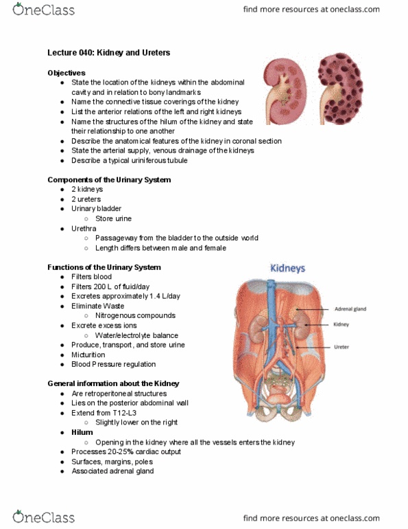
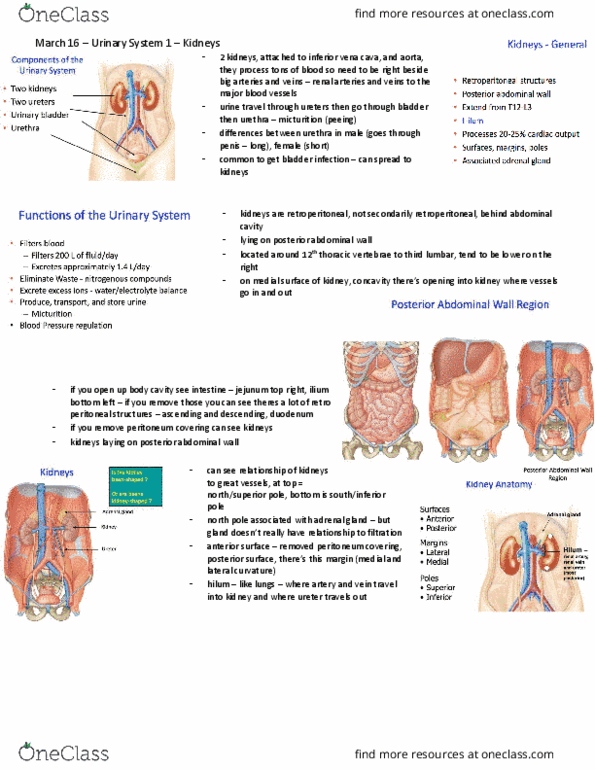
Anatomy Kidneys Bilateral retroperitoneal organs located in posterior abdomen (T12 -
L3)
Right kidney is slightly lower than left kidney (due to liver)
Main function
Filter and excrete waste products (metabolites, excess
electrolytes) from blood
Water and electrolyte balance
External layers
D
eep to superficial
R
enal capsule (tough
fibrous capsule)
P
erirenal fat
(collection of
extraperitoneal fat)
R
enal fascia (enclose
kidneys and
suprarenal glands)
P
ararenal fat
(posterolateral
aspect of kidney)
Internal structure
Renal
parenchyma has 2 areas
Oute
r cortex & inner medulla
Cort
ex extends into medulla
dividing it into renal
pyramids
Renal
pyramid
Apex
(renal papilla) is associated
with minor calyx (collect
urine from pyramids) and
major calyx (union of
multiple minor calices)
Urin
e passes from minor calyx
-> major calyx -> renal
pelvis -> ureter -> bladder
Renal hilum
Deep
find more resources at oneclass.com
find more resources at oneclass.com

fissure in medial margin of
each kidney
Facili
tates entry and exit of renal
vessels & ureter
Blood supply
Arterial Renal arteries (off abdominal aorta L2)
R renal artery crosses posterior to
IVC
L renal artery doesn't cross
anything (shorter)
Segmental arteries
Interlobar arteries (either side of renal
pyramid)
Arcuate arteries
Interlobular arteries (90* to arcuate
arteries)
Pass through cortex
Afferent arterioles
Glomerulus
Efferent arterioles
Peritubular network (supply outer 2/3
cortex) and vasa recta (supply inner 1/3 cortex + medulla)
Venous Renal veins
R renal vein doesn't cross
anything (shorter)
L renal vein crosses anterior to
abdominal aorta
Lymphatics Lateral aortic nodes
Ureters 25cm bilateral long thick tubes transporting urine from kidney (renal
pelvis) -> bladder
Smooth muscle walls produce peristaltic waves
Neurovascular supply
Abdominal part Renal artery & vein
Testicular/ ovarian artery &
vesin
Pelvic part Superior and inferior vesical
arteries & veins
Nervous supply Renal, testicular/ ovarian and
hypogastric plexuses
Sensory supply - T11 to L2
Bladder Main function
Collection, temporary storage (up to 600mL) and expulsion of
urine
Structure
find more resources at oneclass.com
find more resources at oneclass.com

Apex -
connected to umbilicus via
median umbilical ligament
Body
Fundu
s/ base
Neck -
convergence of fundus &
2x inferolateral surfaces
Detru
sor muscle - smooth
muscle in 3 orientations
that receives both SNS and
PSNS innervation
Intern
al urethral sphincter
Male - circular
smooth muscle,
autonomic control
Female - functional
sphincter via bladder
neck and proximal
urethra
Exter
nal urethral sphincter
Skeletal muscle,
voluntary control,
relaxation allows
urine flow
Neurovascular
Arterial Superior vesical branch (internal iliac) +
inferior vesical (M) or vaginal (F)
Obturator and inferior gluteal arteries -
smaller supply
Venous Vesical venous plexus (empty into internal
iliac vein)
Nervous SNS - hypogastric nerve (T12-L2), relaxation
of detrusor muscle, urine retention
PSNS - pelvic nerve (S2-S4), contraction of
detrusor muscle, micturition
Somatic (voluntary) - external urethral
sphincter via pudendal nerve (S2-S4) for constriction (storage) or
relaxation (micturition)
Normal bladder stretch reflex arc (infants)
Sensory afferent nerves to brain in
bladder wall, signaling need to urinate when bladder is full
Bladder wall distends as it fills with
urine; sensory nerves transmit signals to spinal cord
Interneurons in spinal cord signal to
PSNS efferents (pelvic nerve) -> contract detrusor muscle for
micturition
This becomes non-functional after
infancy when voluntary control takes over unless in spinal
injury (descending inhibition cannot reach bladder) or
find more resources at oneclass.com
find more resources at oneclass.com
Document Summary
Outline the anatomy of the urogenital tract including blood supply and lymphatic. Form a differential diagnosis and outline the approach to investigation for macroscopic haematuria. Discuss symptoms that may signify urinary tract trauma and the investigations appropriate to discover the injury. Bilateral retroperitoneal organs located in posterior abdomen (t12 - Right kidney is slightly lower than left kidney (due to liver) Filter and excrete waste products (metabolites, excess electrolytes) from blood. P eep to superficial enal capsule (tough fibrous capsule) erirenal fat (collection of extraperitoneal fat) enal fascia (enclose kidneys and suprarenal glands) ararenal fat (posterolateral aspect of kidney) Cort ex extends into medulla dividing it into renal pyramids pyramid. Apex (renal papilla) is associated with minor calyx (collect urine from pyramids) and major calyx (union of multiple minor calices) > major calyx -> renal pelvis -> ureter -> bladder. Deep fissure in medial margin of each kidney. Facili tates entry and exit of renal vessels & ureter.