PHYL2002 Study Guide - Final Guide: Acetylcholine, Stroke Volume, Endoplasmic Reticulum


Cardiac Muscle Physiology
• Structure
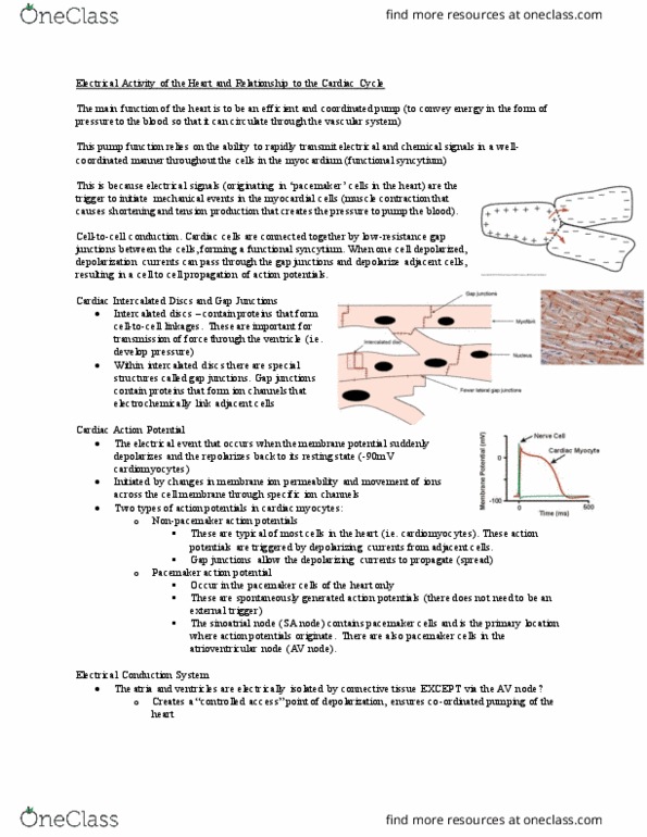
o Intercalated discs join cardiac muscle cells
▪ Desmosomes → connect intermediate filaments → connect
cardiac cells
▪ Tight junctions → stop para-cellular transport
▪ Gap junctions → connect cytosol of 2 cells → allow action
potentials to pass cell to cell
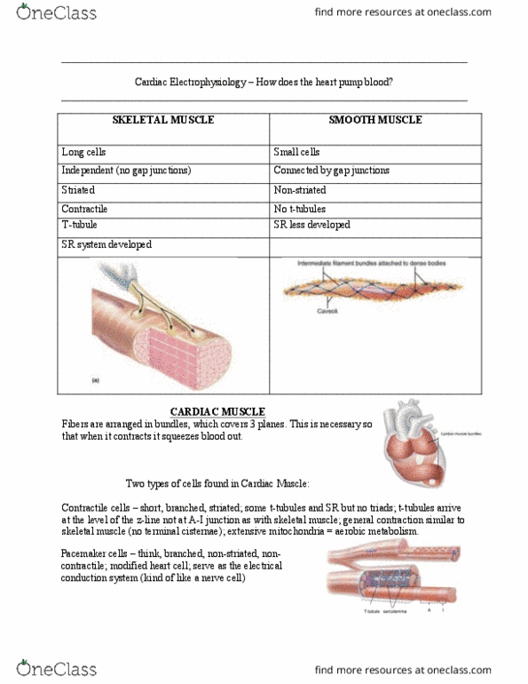
o Many mitochondria
o Sarcomas not perfectly aligned → less organized
o Lots of branching
o Striated
o One nucleolus per cell
o Gastric and intestinal smooth muscle is electromechanically
coupled
o Interstitial cells of Cajal are the pacemaker cells that trigger
depolarization
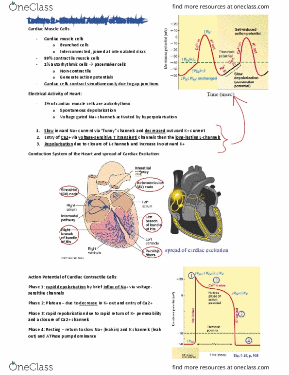
• Cardiac electrical activity
o Pacemaker in SA node generates action potentials
o Action potentials spread cell-to-cell through gap junctions
o Ventricular action potential long (250ms), resting potential
~90mV
o Pacemaker potential spontaneously depolarizes
o Generates rhythmicity
o Action potential → phase 0 → Ca2+ influx, not Na+
o Repolarziation → phase 3 → K+ outflow
o Pacemaker due to fall in K+ outflow
o Increase in Na+ inflow → If → funny current
o Some Ca2+ inflow
o Resting membrane potential of pacemaker cells → not stable
find more resources at oneclass.com
find more resources at oneclass.com