SSEH3301 Study Guide - Final Guide: Interatrial Septum, Interventricular Septum, Mitral Valve

20 Jun 2018
School
Department
Course
Professor

Chapter 2: Cardiovascular Disease
Cardiovascular Disease - diseases of the heart/and or blood vessels (coronary heart, stroke)
→ Most common cause of death in Australia (every hour, 5 Australians die from CVD)
Cardiovascular Anatomy & Physiology
1. Role of CV system:
a. Transport oxygen and nutrients
b. Removal of metabolic waste
2. Heart Anatomy
a. Chambers of the Heart→ Right atrium, right ventricle, left atrium and left ventricle
i. The atria are smaller and have thinner and less muscular walls than
ventricles. Atria act as receiving chambers for blood, so they are
connected to the veins that carry blood to the heart.
ii. The ventricles are larger, stronger pumping chambers that send blood
out of the heart. Ventricles are connected to the arteries that carry blood
away from the heart
b. Valves of the Heart → The heart functions by pumping blood both to the lungs
and to systems of body. To prevent blood from flowing backward/regurgitating
back into the heart, a system of one-way valves are present in the heart.
i. Atrioventricular valves (AV) located between atria and ventricles and
only allow blood to flow from atria into the ventricles.
1. Right side is called triscupid valve
2. Left side is called bicuspid valve.
ii. Semilunar valves
1. Right side is called pulmonary valve prevents backflow from
pulmonary trunk into right ventricle
2. Left side is called aortic valve prevents backflow from aorta into
left ventricle
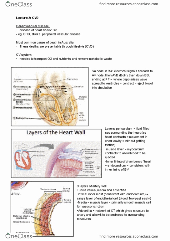
3. Electrical Activity of the Heart
a. Heart is able to set its own rhythm and conduct signal. The cardiac muscle cells
in heart are responsible to set pace for the for cardiac muscle cells.
b. Conduction system
i. SA node → Sinoatrial node (pace of heart as a whole and signals atria to
contract)
ii. AV node → Atrioventricular node (picks up signal sent by SA node and
transmits thoough AV bundle/bundle of his)
iii. AV Bundle/Bundle of His → Strand of connective tissue that runs
through the interatrial septum and into the interventricular septum.
iv. Bundle branches → AV bundle splits into left and right branches running
through the septum until reaching apex of heart.

v. Purkinje Fibres → Branching off from left and right bundle branches are
many Purkinje fibers that carry signal to walls of ventricles, stimulating
cardiac muscle cells to contract and pump blood out of the heart.
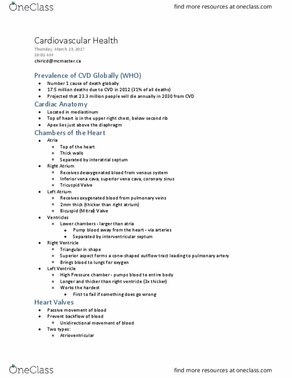
4. Layers of the Heart Wall → 3 layers
a. Epicardium/Visceral layer of pericardium
i. Outermost layer of heart wall
ii. Helps to lubricate and protect the outside of the heart.
b. Myocardium
i. Muscular middle layer of the heart wall containing cardiac muscle
tissue.
c. Endocardium
i. Innermost layer of heart wall
ii. Very smooth and is responsible for keeping blood from sticking
inside of heart and forming potentially deadly blood clots.
Thickness of heart wall varies in different parts of the heart.
5. The Normal Artery
a. Tunica Intima (inner most layer)
b. Tunica Media (muscle layer of artery - smooth muscle)
c. Tunica Adventitia (network of connective tissue)
6. Atherosclerosis
a. Disease causing thickening and loss of elasticity of arterial walls
b. Characterized by formation of lipid and high concentration of cholesterol in intima
or media of large and medium-sized arteries (build up of fat in intima or media)
c. Causing complete blockage of vessel
d. Primarily affects → coronary, illiac, femoral, aorta
→ AFFECTS Heart circulation
e. Creating blockages → not enough blood to get through to transport oxygen and
nutrients
Pathogenesis of Atherosclerosis
→ Start with endothelium (barrier between blood and artery wall, selectively permeable,
produce hormones and other substance, has receptors)
→ Endothelial injury due to:
1. Blood-borne chemicals (tobacco, cholesterol LDL, high blood glucose)
2. Hypertension
3. Vasoconstrictor substances
4. Infections
Document Summary
Cardiovascular disease - diseases of the heart/and or blood vessels (coronary heart, stroke) Most common cause of death in australia (every hour, 5 australians die from cvd) Cardiovascular anatomy & physiology: role of cv system, transport oxygen and nutrients, removal of metabolic waste, heart anatomy, chambers of the heart right atrium, right ventricle, left atrium and left ventricle i. ii. The atria are smaller and have thinner and less muscular walls than ventricles. Atria act as receiving chambers for blood, so they are connected to the veins that carry blood to the heart. The ventricles are larger, stronger pumping chambers that send blood out of the heart. Ventricles are connected to the arteries that carry blood away from the heart: valves of the heart the heart functions by pumping blood both to the lungs and to systems of body. To prevent blood from flowing backward/regurgitating back into the heart, a system of one-way valves are present in the heart.