BIOC 4580 Study Guide - Midterm Guide: Oligosaccharide, Speedstep, Amphiphile

12 Feb 2017
School
Department
Course
Professor
Document Summary
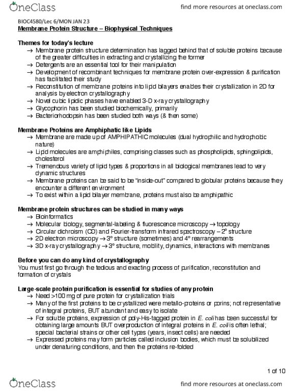
Membrane protein structures are less studied than soluble proteins because of difficulties extracting and crystallization. Reconstitution of membrane proteins into lipid bilayers enables their crystallization in 2 dimensions for analysis by electron crystallography. Novel cubic lipidic phases have enabled 3d x-ray crystallography. Membrane proteins are amphipathic (cid:862)i(cid:374)side-out(cid:863) compared to globular proteins because they encounter a different environment. Molecular biology, segment-labeling, and fluorescence microscopy topology. Circular dichroism (cd) and fourier-transform infrared spectroscopy secondary structure. 2d electron microscopy tertiary (sometime quaternary) structure. Epr and nmr spectroscopy tertiary structure, mobility, dynamics, interactions with membranes. Add p(cid:396)e(cid:272)ipita(cid:374)ts (cid:894)to se(cid:395)ueste(cid:396) (cid:449)ate(cid:396) (cid:373)ole(cid:272)ules a(cid:374)d a(cid:272)hie(cid:448)e a slo(cid:449) (cid:862)salti(cid:374)g out(cid:863)(cid:895) Add protein stabilizers (eg. sugars, cations, cofactors) Nucleation of a few sites for crystal formation. Metastable zone protein micelles aggregate and dissociates until supersaturation and protein forms crystal. Membrane protein crystallography lags behind soluble protein crystallography. Intensive screening trials must be done for crystallization conditions of a water-insoluble protein.