BIOC 4580 Study Guide - Midterm Guide: Actomyosin Ring, Fluorescence Microscope, Intermediate Filament

6 Mar 2017
School
Department
Course
Professor

Document Summary
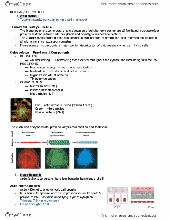
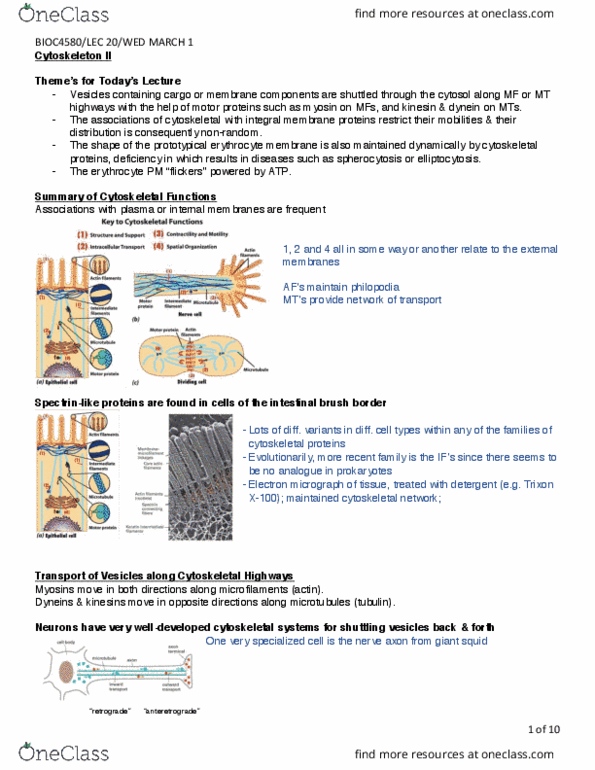
The biogenesis, shape, structure, and dynamics of cellular membranes are all facilitated by cytoskeletal proteins that can interact with peripheral and/or integral membrane proteins. The 3 major cytoskeletal protein families are microfilaments, microtubules, and intermediate filaments, all with a variety of accessory proteins. Fluorescence microscopy is a major tool for visualization of cytoskeletal dynamics in living cells. An interlocking 3-dimensional scaffolding that extends throughout the cytosol and interfacing with the plasma membrane. Modulation of cell shape and cell movement. The 3 families of cytoskeletal proteins vary in composition and thickness: microfilaments. Actin (eukaryotic protein; there is a bacterial homologue mreb) Actin ~10% of total eukaryotic cell protein: mfs (cid:271)ou(cid:374)d to specific membrane proteins lie just beneath & parallel to pm cortex is underlying layer of cytoplasm. Major part is the cortical actin maintains cell structure and shape. G-actin (globular) 42 kda monomer exists free at low ionic strength. F-actin (filamentous) at physiological ionic strength (double helix of monomers)