HK 3401 Study Guide - Midterm Guide: Flexor Digitorum Superficialis Muscle, Pronator Teres Muscle, Brachial Artery

22 Oct 2016
School
Department
Course
Professor
Document Summary
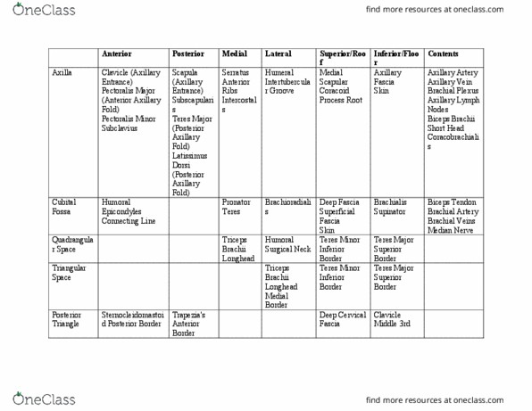
Cubital fossa: shallow triangular depression on anterior surface of the elbow, boundaries: Superior: imaginary line that connect medial and lateral epicondyles. Floor: formed by brachialis and supinator muscle. Roof: formed by brachial and antebrachial fascia, reinforced by bicipital aponeurosis, subcutaneous tissue and skin: contents of cubital fossa: Terminal part of brachial artery and the beginning of radial and ulnar arteries that are terminal branches from the brachial artery. Radial nerve, dividing into superficial and deep branches. Forearm contains 2 bones, the ulna and the radius, joined together by the interosseous membrane. Forearm role is to assist shoulder in application of force and controlling placement of hand in space. Flexor and pronator muscles of forearm are in the anterior compartment of the forearm (mostly innervated by the median nerve with 1. 5 exceptions) The extensors and supinators are in the posterior compartment (innervated by radial nerve)