PSYC 2410 Study Guide - Cognitive Neuroscience, Neurochemistry, Frontal Lobe

6 Apr 2013
School
Department
Course
Professor
Document Summary
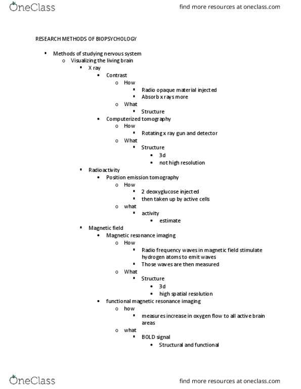
5. 1 methods of visualizing and stimulating the living human brain. Conventional x-rays: based on the fact that different structures absorb x-rays differently which shows up on film. It works when there is a high contract between the tissues being examined (ex: bones vs soft tissue). It"s not good for brain scans because the different tissues of the brain have similar x-ray absorption. Contrast x-rays: a contrast dye is injected and is photographed", shows blood vessels usually. X-ray computed tomography (ct): a rotating x-ray tube takes multiple 2d images which are combined to create 3d images. Magnetic resonance imaging (mri): radio frequency waves align hydrogen in water molecules which then emit a measurable magnetic signal; creates both 2d and 3d images and is clearer than ct. Positron emission tomography (pet): a radioactive compound is injected (2-deoxyglucose or 2-dg) which accumulates in active cells and degrades slowly. The radioactive signal of active cells is greater than that of non-active cells.