NURS 1750 Study Guide - Midterm Guide: Cerebral Circulation, Chemotherapy, Palpitations

28 Jun 2018
School
Department
Course
Professor

Date of Midterm: November 15, 2017
Anatomy and Physiology Midterm Study Guide #2
Questions/Answers:
Week Six (Chapter 13 and 16)
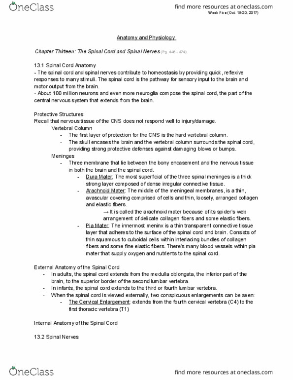
1. Name the three protective layers of the CNS in order.
a. Hard boney skull and vertebral column
b. Meninges (connective tissue)
c. Cerebrospinal fluid
2. Name and describe the three meninges in order from superficial to deep.
a. Dura Mater:
a.i. Dense irregular connective tissue.
a.ii. Forms a sac from the level of the foramen magnum in the occipital bone
where it is continuous with the meningeal dura mater of the brain.
a.iii. Continuous with outer covering of spinal and cranial nerves
a.iv. Most superficial.
b. Arachnoid Mater:
b.i. Continuous through the foramen magnum into the brain.
b.ii. Avascular covering- cells & collagen and elastic fibers.
b.iii. Named because the mater resembles a spider’s web.
b.iv. Middle of the meningeal membrane.
c. Pia Mater:
c.i. Connective tissue layer-adheres to the surface of the spinal cord and
brain.
c.ii. Squamous to cuboidal cells, collagen & elastic fibers
blood vessels supply oxygen and nutrients to the spinal cord.
c.iii. Innermost layer.
c.iv. Suspend the spinal cord in the middle of its dural sheath (denticulate
ligaments)-protect against displacement.
3. What is the subdural and subarachnoid space?
a. Subdural Space: Space between dura mater and arachnoid mater. Contains
interstitial fluid.
b. Subarachnoid Space: between the arachnoid mater and pia mater. Contains
shock absorbing cerebrospinal fluid.
4. Describe the procedure of a spinal tap, the location and give a few reasons why its
preformed.
a. A local anesthetic is given, and a long, hollow needle is inserted into the
subarachnoid space to withdraw cerebrospinal fluid (CSF).
b. Patient lies on side with the vertebral column flexed.
c. Performed to:
c.i. Introduce antibiotics
c.ii. Contrast media
c.iii. Anesthetics
find more resources at oneclass.com
find more resources at oneclass.com

Date of Midterm: November 15, 2017
c.iv. Chemotherapy
c.v. Measure CSF pressure
c.vi. Evaluate the effects of treatment-meningitis
d. Normally performed in adults between L3 and L4 or L4 and L5 lumbar vertebrae
because this region provides safe access to the subarachnoid space without risk
of damaging the spinal cord (spinal cord ends at vertebra L2; spinal meninges
and cerebrospinal fluid extend to the second sacral vertebra (S2)).
5. In adults, the beginning of the spinal cord extends from the medulla oblongata. In
newborns it extends from the 3rd or 4th lumbar vertebra and stops growing around age 4
or 5, but the vertebral column continues.
6. ???
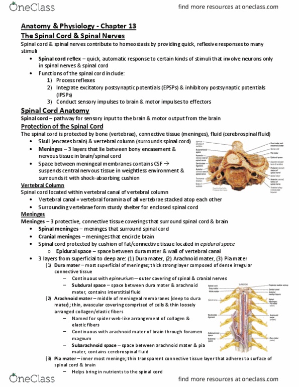
a. Cervical Enlargement: ends from fourth cervical vertebra (C4) to the first thoracic
vertebra (T1). Nerve to and from upper limbs arise from the cervical enlargement.
b. Lumbar Enlargement: Extends from the ninth to the twelfth thoracic vertebra (T9-
T12). Nerves to and from the lower limbs arise from the lumbar enlargement.
c. Filum Terminale: is an extension of pia mater. It fuses with the arachnoid mater
and dura mater, and anchors the spinal cord to the coccyx.
d. Spinal Nerves: are the paths of communication between the spinal cord and
specific regions of the body.
e. Bundles of axons: roots (two kinds), connect spinal nerve to a segment of the
cord by even smaller bundles of axons called rootlets.
f. Posterior (Dorsal) Root Ganglion: cell bodies of sensory neurons.
g. Posterior (Dorsal) Root: contains only sensory axons.
h. Anterior (Ventral) Root: contain axons of motor neurons.
i.
7. Name three functions of the spinal cord.
a. Process reflexes.
b. Integrate (gray matter) postsynaptic potentials.
c. Conduct sensory impulses to brain - motor impulses to effectors.
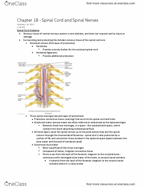
8. Name some differences between grey and white matter.
White Matter Grey Matter
find more resources at oneclass.com
find more resources at oneclass.com

Date of Midterm: November 15, 2017
Gray Matter Horns:
Spinal Cord and Brain
Clusters of neuronal cell bodies-(functional
groups called nuclei)
Sensory nuclei receive input from receptors
via sensory neurons
Motor nuclei provide output to effector tissues
via motor neurons
White matter columns-
Bundles of axons: travel up & down spinal
cord- called tracts.
Tracts are bundles of axons-CNS
Nerves are bundles of axons- PNS
Sensory (ascending) tracts: axons- nerve
impulses toward the brain
Motor (descending) tracts: axons- nerve
impulses from the brain
9. Upper Motor Neuron affects brain and spinal cord. Lower Motor Neuron affects spinal
cord (brainstem) and skeletal muscles.
10. Define what sensory and motor tracts consist of:
a. Sensory (ascending/afferent): tracts consist of axons that conduct nerve
impulses toward the brain.
b. Motor (descending/efferent): tracts consist of axons that carry nerve impulses
from the brain
11. Sensory and motor tracts of the spinal cord are continuous with sensory and motor tracts
in the brain.
12. Sensory receptors detect a sensory stimulus.
13. Sensory neurons go to the nerve impulse, which go to the spinal nerve then to the
posterior root and finally to one of three paths. Name each path:
a. White matter of spinal cord →brain as sensory tract
b. Posterior gray horn→ interneurons→ brain as sensory tract
c. Posterior gray horn→ interneurons→ somatic motor neurons involved in spinal
reflex pathways.
14. Give the pathway of motor and sensory tracts
a. Motor:
a.i. Spinal Cord → Motor Output → Skeletal Muscles
a.i.1. Brain → White matter → Somatic Motor Neurons (directly
or indirectly; neurons).
b. Sensory:
b.i. Spinal cord → Motor output → cardiac muscle, smooth muscle & glands
b.i.1. Brain→ lateral gray horn-→ anterior gray horn→ anterior
root →spinal nerve →autonomic motor neuron
15. Give the pathway of autonomic and somatic motor neurons:
a. Autonomic motor neuron → autonomic motor neurons in peripheral nervous
system (PNS) → cardiac muscle, smooth muscle & glands (effector).
b. Somatic Motor Neurons: motor output (nerve impulses) → anterior gray horn →
anterior root → spinal nerve → skeletal muscles (effector).
16. There are 31 pairs of spinal nerves. They emerge at regular intervals from intervertebral
find more resources at oneclass.com
find more resources at oneclass.com
Document Summary
Forms a sac from the level of the foramen magnum in the occipital bone where it is continuous with the meningeal dura mater of the brain. Continuous with outer covering of spinal and cranial nerves a. iii. a. iv. Continuous through the foramen magnum into the brain. Avascular covering- cells & collagen and elastic fibers. Named because the mater resembles a spider"s web. b. i. b. ii. b. iii. b. iv. Middle of the meningeal membrane: pia mater: c. i. c. ii. c. iii. c. iv. Connective tissue layer-adheres to the surface of the spinal cord and brain. Squamous to cuboidal cells, collagen & elastic fibers blood vessels supply oxygen and nutrients to the spinal cord. Suspend the spinal cord in the middle of its dural sheath (denticulate ligaments)-protect against displacement: what is the subdural and subarachnoid space, subdural space: space between dura mater and arachnoid mater. Contains interstitial fluid: subarachnoid space: between the arachnoid mater and pia mater.