Anatomy and Cell Biology 3309 Study Guide - Final Guide: Stratified Squamous Epithelium, Muscular Layer, Muscularis Mucosae

22 May 2018
School
Department
Professor

Histology 3309
Lab 16
Digestive System II
Identify the papillae. What glands are found at the base of this papillae?
- The papillae is circumvallae papilla
- Von Ebner’s glands
Identify the salivary gland
- Parotid gland
- No mucous
- Only serous
Following is a section of parotid gland. Identify the duct shown
- Striated duct
o Has basal striations (invaginations of the plasma at the bottom of the nucleus)
find more resources at oneclass.com
find more resources at oneclass.com

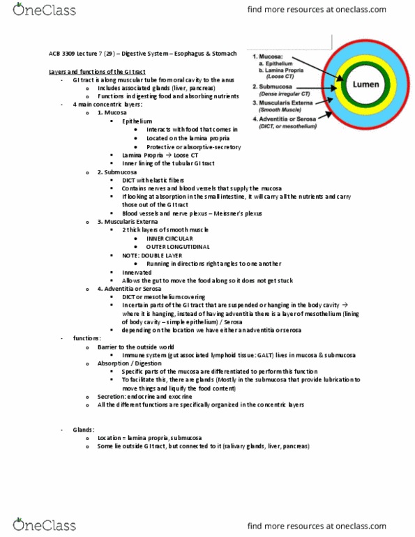
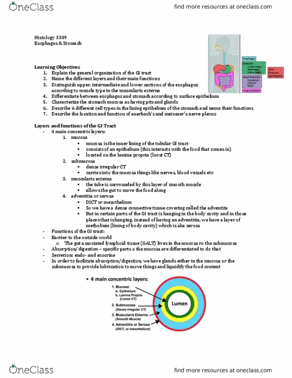
- Mucosa
o Has epithelium
o Lamina propria
o Muscularis mucosa
- Submucosa
o Made of DICT but has special glands in the esophagus and duodenum
- Muscularis externa
o Mainly made of 2 layers of smooth musclce: the inner circular layer and the outer
longitudinal layer
o Has unique features in esophagus and stomach
- Adventitia or serosa
o Blends with the outer connective tissue
Esophagus
- This is a cross section of the esophagus
- The wall has a lining of stratified squamous epithelium (blue arrow), which is non-
keratinizing in humans
- The lamina propria sends a number of projections into the epithelium
- The muscularis mucosae (yellow arrow) contains typical smooth muscle cells
find more resources at oneclass.com
find more resources at oneclass.com

- This is a cross section of the esophagus
- Right at the center, you see the lumen of the esophagus
- Identifying them from the inner to the outer site,
- M = mucosa
- Blue arrow: mucosa epithelium
o Epithelium of the esophagus is the stratified squamous
▪ Stratified squamous epithelium is present in places wehre there is a great
deal of wear and tear (like skin – but the skin is keratinized)
o Esophagus epithelium is non keratinized
- Bw the blue and yellow arrow: lamina propria
o Loose CT
o Contains reticular cells and lymphocytes
- Yellow arrow: muscularis mucosa
o Thin layer of smooth muscle
- Green arrow: submucosa (SM)
o Contains blood vessels and lymphatics
o Contains glands called esophageal glands
- Orange box: muscularis externa (ME)
o Has inner circular layer (fibers run in a circular fashion)
o And
- Muscularis externa (zoomed into the orange box)
- Has inner circular layers (fibers run in circular fashion) and outer longitudinal layer (fibers
run from top to bottom, in straight line)
- What is special of muscularis externa of esophagus?
o The upper 1/3 of esophagus consists of both these layer (inner circular and outer
longitudinal) or striated muscule (aka skeletal muscle)
o The middle 1/3 of esophagus consists of smooth and skeletal muscle
o The lower 1/3 consists of only smooth muscle
▪ From there one, there is only smooth muscle in the GI tract
find more resources at oneclass.com
find more resources at oneclass.com
Document Summary
Striated duct: has basal striations (invaginations of the plasma at the bottom of the nucleus) Mucosa: has epithelium, lamina propria, muscularis mucosa. Submucosa: made of dict but has special glands in the esophagus and duodenum. Muscularis externa: mainly made of 2 layers of smooth musclce: the inner circular layer and the outer longitudinal layer, has unique features in esophagus and stomach. Adventitia or serosa: blends with the outer connective tissue. The wall has a lining of stratified squamous epithelium (blue arrow), which is non- keratinizing in humans. The lamina propria sends a number of projections into the epithelium. The muscularis mucosae (yellow arrow) contains typical smooth muscle cells. This is a cross section of the esophagus. Right at the center, you see the lumen of the esophagus. Identifying them from the inner to the outer site, Bw the blue and yellow arrow: lamina propria: loose ct, contains reticular cells and lymphocytes. Yellow arrow: muscularis mucosa: thin layer of smooth muscle.