BICD 110 Midterm: Midterm Study Guide

28 Jun 2018
School
Department
Course
Professor


BICD110 Midterm Study Guide
Lecture 1
- Microscopy: Cell is below limit of resolution of human eye. Use microscopy to visualize cells
oHuman eye can see 100 µm object. Average animal cell is 10-20 µm in diameter
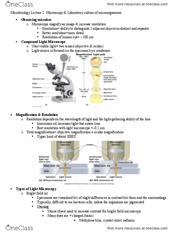
obasic format of light microscope
- 2 factors important in whether an object can be seen
o1) magnification
o2) resolution
limit of resolution: minimum distance 2 objects approach another and still appear
separate
Limit of resolution for light microscope is ½of light. For visible light, = 0.4 – 0.7 µm.
½ = 0.2 – 0.35 µm, so objects bigger than this will affect light and be visible, and
small objects will not be visible because no perturbation of waves will occur
Smallest objects visible with a light microscope are mitochondria and small bacteria,
which is 400x better than the naked eye
- problems of microscopy
o1) cells are 70% H2O, so not much detail can be seen
solution: use stains and dyes for different components of the cell, like DNA, proteins,
membranes, etc. These stains and dyes can bind to the components and refract light
o2) cells are fragile. They become distorted or broken by observation
solution: cells fixed before observation to become stronger
methanol fixation: causes proteins to denature, precipitate out in place, and
complex to each other
chemical crosslinkers: formaldehyde or
glutaraldehyde used; interact with lysines in
proteins to complex together, allowing protein stabilization
o3) tissues are thick and light can’t penetrate
solution: embed tissue in something stronger than the tissue (like paraffin wax),
then slice into sections with microtome to allow 1-10 µm thick slices of tissue
o4) cells are killed by staining, fixing, and sectioning.
Possible you’re looking at an artifact (artificial fact
because the cells are dead and look different)
solution: confirm results by looking at living cells
find more resources at oneclass.com
find more resources at oneclass.com

interference microscopy (phase contrast microscopy): used to look at living cells
with light instead of staining/dying/fixing. As light passes from less
dense to more dense material, its speed slows. The change in speed
is used to enhance contrast in living unstained cells
- Electron microscopy: used to observe objects smaller than 0.2 – 0.35 µm.
When electrons are accelerated, they have a characteristic wavelength.
Electrons through 50,000 V acceleration have wavelength of 0.005 nm
oLimit of resolution should be ½ wavelength = 0.0025 nm. However, real
limit of resolution is 0.1 nm (still 400x better than light microscopy)
oStandard transmission EM (TEM): 50,000 – 100,000 V; 2D
Under right circumstances, can see DNA, ribosomes, and proteins.
Same problems require fixing, embedding, sectioning, and staining
Fix -> embed in plastic (very hard) -> section with diamond knife (50 – 100 nm
sections) -> stain with heavy metal solution (lead, uranium, osmium) to make
objects they bind to dense
Can also stain with spray of heavy metal, called negative staining
oScanning EM (SEM): 3D
Put sample on grid -> coat with platinum -> use scanning EM machine to detect
Electron gun shoots electrons at coated sample. Detector moves around
sample and collects 2° electrons that have deflected off the platinum coat,
integrates them, and processed them into 3D picture on screen
Can magnify 20,000x. Good for fly eyes, bristles, pollen, and stereocilia on
cells in your ears
oHigh resolution scanning EM (FEISEM): 3D
Can magnify 100,000x. Good for seeing single microtubule or nuclear pore
oFreeze fracture EM: 3D
freeze cell in liquid nitrogen (-196°C) -> fracture with knife -> coat
with metal -> view
Line of fracture is where the least amount of resistance occurs, like
in middle of membrane to expose proteins
oFreeze etch EM: 3D with better resolution
Freeze cells in liquid helium (-269°C) -> fracture with knife -> etch
away H2O with vacuum -> coat with metal -> view
Can see more details since less water is there to hinder resolution
Excellent preservation, no artifacts, and lots of details
oCyro electron tomography EM: 3D, 1 nm resolution (best resolution)
- Immunofluorescence (IF)
oUse antibodies as specific stains
oAntibodies produced normally by vertebrates as a defense against infection. Millions of
different ones are produced, each recognizing different molecules or antigens
oInject protein of interest into rabbit, to produce lots of antibodies to specific protein.
Fluorescently label antibody to achieve a specific stain
find more resources at oneclass.com
find more resources at oneclass.com

oFix cells (either as a section or grown on coverslip) -> permeabilize the
cells (with mild detergent) -> add fluorescently-labeled antibody (ex:
anti-tubulin) -> let it bind to its structure (ex: microtubules) -> wash
away excess antibody -> look at fluorescence under the microscope
oAllows you to do the following:
Determine the presence or absence of a protein in a cell
Determine the location and pattern of protein in cell
Determine the quantity of the protein
Determine how the above are altered in the cell cycle, development, and disease
oGFP-labeling of live organisms
Gene for GFP can be fused to gene of interest, then transfected back into
cell line or even into a whole organism
Gene can also be engineered to be expressed only in desired tissue or cell
type (ex: red blood cells, white blood cells, heart, brain, wing, etc.)
Can look at fixed/live cells, live organisms, or take movies of live organisms
oDr. Roger Tsien: developed many different GFP mutants to label
different cell parts with different colors
Lecture 2
- Cell separated from environment by membranes. Membranes protect from environment
and isolate specific cellular functions from one another. Bring order to the cell
- Membranes are very fluid
oLaser tweezers: can point lasers to pull at part of membrane to bring them apart, but
not break them. The membrane components move quickly to adapt to the distortion of
the layers. Only things that hold membranes together are non-covalent interactions.
Can look at mitosis and pull chromosome off spindle to study
- All biological membranes are:
oMade of lipids and proteins
oHeld together by non-covalent bonds
- Properties of membranes
oBarrier: to isolate processes
Lipids provide barrier and flexibility of barrier
Proteins provide selectivity
oA rigid structure, but flexible and dynamic
- Typical cell’s plasma membrane: 50% lipids and 50% proteins by mass
oMany more lipids (109), because lipids are smaller in size
- Bilayers
oEarly theoretical models for membrane structure
oBasic structure of cell membranes: fluid mosaic model (John Sanger)
Different parts put together to make a whole. Fluid because proteins and lipids can
move around through the membrane
oSupporting evidence for bilayer
find more resources at oneclass.com
find more resources at oneclass.com
Document Summary
Microscopy: cell is below limit of resolution of human eye. Use microscopy to visualize cells: human eye can see 100 m object. Average animal cell is 10-20 m in diameter: basic format of light microscope. 2 factors important in whether an object can be seen: 1) magnification, 2) resolution limit of resolution: minimum distance 2 objects approach another and still appear separate. Limit of resolution for light microscope is of light. For visible light, = 0. 4 0. 7 m. = 0. 2 0. 35 m, so objects bigger than this will affect light and be visible, and small objects will not be visible because no perturbation of waves will occur. These stains and dyes can bind to the components and refract light: 2) cells are fragile. They become distorted or broken by observation solution: cells fixed before observation to become stronger. As light passes from less dense to more dense material, its speed slows.