PSY2061 Chapter Notes - Chapter 5: Transcranial Magnetic Stimulation, Operant Conditioning, Default Mode Network

5 Jun 2018
School
Department
Course
Professor

PSY2061 – Readings – Week 2
The research methods of biopsychology
~ methods of studying the nervous system
• methods of visualising or stimulating the living human brain
•
o x-ray based techniques
o
▪ an x-ray beam is passed through an object and then onto a
photographic plate
▪ each of the molecules through which the beam passes
absorbs some of the radiation, thus only the unabsorbed
portions of the beam reach the photographic plate
▪ effective in characterising internal structures that differ
from their surroundings
▪ carries little information about brain structures
▪ contrast x-rays
▪
▪ involve injecting a substance into one compartment
of the body that absorbs x-rays either less or more
than the surrounding tissue
▪ cerebral angiography
▪
▪ use the infusion of a radio-opaque dye into a
cerebral artery to visualise the cerebral
circulatory system during x-ray photography
▪ most useful for localising vascular damage
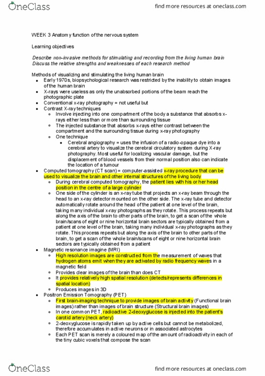
▪ computed tomography
▪
▪ a computer-assisted x-ray procedure that can be
used to visualise the brain and other internal
structures of the living body.
▪ the neurological patient lies with his or her head
positioned in the centre of a large cylinder
▪
▪ on one side of the cylinder is an x-ray tube
that projects an x-ray beam through the head
to an x-ray detector mount on the other side
▪ these rotate around the head - taking many
individual x-ray photographs as they rotate
▪ each x-ray photograph is combined by a
computer to generate a CT scan of one
horizontal section of the brain
▪ process is repeated along another level of the
brain - when combined they provide a 3D
representation of the brain
o radio-activity based techniques
o
▪ positron emission tomography - PET
▪
find more resources at oneclass.com
find more resources at oneclass.com

▪ provides images of brain activity - functional brain
images rather than images of brain structure -
structural brain images
▪ radioactive fluorodeoxyglucose FDG - is injected into
the patient’s carotid artery - cannot be metabolised -
therefore accumulate in active neurons or in
associated astrocytes - until it is gradually broken
down
▪ resulting scan will indicate the areas of the target
brain level that were most active
▪ each scan is a coloured map of the amount of
radioactivity in each of the tiny cubic voxels -
volume pixels that compose the scan
▪ use in identifying the distribution in the brain of
molecules of interest
▪
▪ injecting volunteers with radioactively
labelled ligands - ions or molecules that bind
to other molecules under investigation
▪ magnetic-field-based techniques
▪
▪ structural brain-imaging procedure in which high
resolution images are constructed from the
measurement of radio-frequency waves that
hydrogen atoms emit as they align with a powerful
magnetic field
▪ provides relatively high spatial resolution - the
ability to detect and represent differences in specific
location
▪ MRI can produce images in three dimensions
▪ functional MRI
▪
▪ widely used for diagnosis
▪ functional MRI - fmri
▪ produces images representing the increase in
oxygen flow in the blood to active areas of the
brain
▪ active areas of the brain take up more
oxygenated blood than they need for energy
requirements - thus oxygenated blood
accumulates in active areas of the brain
▪ oxygenated blood has magnetic properties
that influence the radio-frequency waves
emitted by hydrogen atoms in an MRI
▪ the signal recorded by fMRI is called the
BOLD signal - the blood-oxygen-level-
dependent signal
▪ Functional MRI has four advantages over
PET: (1) Nothing has to be injected into the
find more resources at oneclass.com
find more resources at oneclass.com

volunteer; (2) it provides both structural and
functional information in the same image; (3)
its spatial resolution is better; and (4) it can
be used to produce three-dimensional images
of activity over the entire brain.
▪ has poor temporal resolution - poor at
specifying the timing of neural events
▪ diffusion tensor imaging
▪
▪ a method of identifying those pathways along which
water molecules rapidly diffuse
▪ provides an image of major tracts - tracts - bundles
of axons - are the major routes of water diffusion in
the brain
▪ important to understand the connections between
structures - connectome
o transcranial stimulation
o
▪ a way of turning off particular areas of cortex
▪ also can turn on areas of cortex
▪ transcranial magnetic stimulation - TMS -
▪
▪ is a technique that can be used to turn off an area of
human cortex by creating a magnetic field under a
coil positioned next to the skull
▪ the magnetic stimulation temporarily turns off part
of the brain while the effects of the disruption
on cognition and behaviour are assessed
▪ often employed to circumvent the difficulty
in determining causation
▪ transcranial direct current stimulation - tDCS
▪
▪ technique used to stimulate - turn on an area of the
cortex by applying an electrical current through two
electrodes placed directly on the scalp
▪ the electrical stimulation temporarily increases
activity in part of the brain while the effects of the
stimulation on cognition and behaviour are
assessed
~ recording human psychophysiological activity
• methods of recording physiological activity from the surface of the human
body
• psychophysiological measures of brain activity
•
o scan electroencephalography - EEG
o
▪ a measure of the gross electrical activity of the brain
▪ recorded through large electrodes by a device called an
EEG
find more resources at oneclass.com
find more resources at oneclass.com