LQB185 Chapter Notes - Chapter 20: Heart Valve, Papillary Muscle, Pulmonary Circulation

25 May 2018
School
Department
Course
Professor

20.2 Heart Valves and Circulation of Blood
➢ As each chamber of the heart contracts, it pushes a volume of blood into
a ventricle or out of the heart into an artery.
➢ Valves open and close in response to pressure changes as the heart
contracts and relaxes.
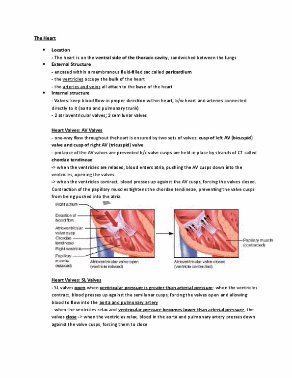
➢ Operation of the Atrioventricular Valves (AV)
- Tricuspid and Bicuspid valves make up the atrioventricular valves
- When the AV is open the rounded ends of the cusps project into
the ventricle (inwardly sucked onwards). Subsequently the
ventricles are relaxed, the papillary muscles are relaxed making
the chordae tendinous go slack and blood moves from a higher
pressure in the atria to a lower pressure in the ventricles
- When ventricles contract, the pressure of the blood drives the
cusps upward until their edges meet and close the opening. At
the same time the papillary muscles contract which pulls on and
tightens the chordae tendinous. This prevents the valve cusps
from everting (opening).
➢ Operation of the Semilunar Valves (SL)
- Aortic and Pulmonary valves make up the Semilunar valves
- Each oes cusps are split ito flaps. The AV ol hae flaps
- SL valves allow ejection of blood from the heart into arteries but
prevent backflow of blood into ventricles.
- Cusps project into the lumen of the artery
- When ventricles contract, pressure builds inside chamber thus
stimulating the valves to open.
find more resources at oneclass.com
find more resources at oneclass.com

- When the ventricles relax, blood starts to backflow toward heart.
Blood fills the valve cusps causing the free edges of the SL to
contract and close entry.
➢ Systemic and Pulmonary Circulations
- Input of one becomes the output of the other
- Left side of the heart is the pump for systemic circulation:
receives red oxygenated blood from the lungs.
- Right side of the heart is the pumps for pulmonary circulation:
receives all the dark-red deoxygenated blood returning from the
systemic circulation.
find more resources at oneclass.com
find more resources at oneclass.com

➢ Coronary Circulation
- Nutrients are not able to diffuse in the heart.
- The Myocardium has its own network of blood vessels (the
Coronary circulation).
- Coronary Arteries branch from the ascending aorta and encircle
the heart
- When heart is contracting, blood flows in the coronary arteries is
squeezed shut then when the heart relaxes the high pressure of
blood propels blood through the coronary arteries, into capillaries
and into coronary veins.
➢ Coronary Arteries
- Left and right arteries branch from the ascending aorta and supply
oxygenated blood to the myocardium.
- Left supples anterior interventricular branch
- Right supples posterior interventricular branch
➢ Coronary Veins
- Blood -> arteries of the coronary circulation -> capillaries (delivers
oxygen and nutrients to heart muscles and collects carbon dioxide
and waste) -> coronary veins.
- Deoxygenated blood from myocardium drains into the Coronary
Sinus -> empties into the right atrium.
- Carrying blood into the coronary sinus are:
1. Great cardiac vein
find more resources at oneclass.com
find more resources at oneclass.com
Document Summary
As each chamber of the heart contracts, it pushes a volume of blood into a ventricle or out of the heart into an artery. Valves open and close in response to pressure changes as the heart contracts and relaxes. Tricuspid and bicuspid valves make up the atrioventricular valves. When the av is open the rounded ends of the cusps project into the ventricle (inwardly sucked onwards). Subsequently the ventricles are relaxed, the papillary muscles are relaxed making the chordae tendinous go slack and blood moves from a higher pressure in the atria to a lower pressure in the ventricles. When ventricles contract, the pressure of the blood drives the cusps upward until their edges meet and close the opening. At the same time the papillary muscles contract which pulls on and tightens the chordae tendinous. This prevents the valve cusps from everting (opening). Aortic and pulmonary valves make up the semilunar valves. Each o(cid:374)e(cid:859)s cusps are split i(cid:374)to (cid:1007) flaps.