KINESIOL 1Y03 Chapter Notes - Chapter 13: Cholecystitis, Hyoscine Butylbromide, Pancreatic Pseudocyst

22 Jun 2018
School
Department
Course
Professor

Gastrointestinal applications of CT imaging
2 major advances in CT have been:
1) Spiral CT: can take pictures with CT coil ‘spread out’ computer then intracalates data
2) multislice CT
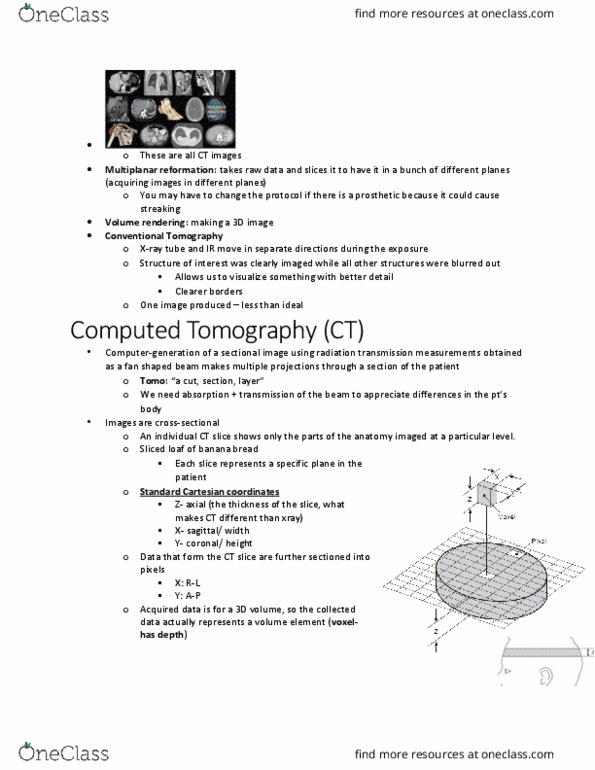
CT images are composed of voxels which is are 3D pixels, giving a measure of slice thickness
This allows scans to be manipulated to produce multiplanar reformants, and 3D reconstructions.
- CT is normally axial but can also have coronal and sagittal reformants
Hounsfield units are the amount of x-ray absorption with reference to water i.e. water =
0HU, air = -1000HU etc.
The Hounsfield scale informs diagnosis by allowing calculation of the HU and then knowing
what density something must be e.g. if pathology is fluid/fat
The naked eye can only see about 10 different levels of ‘grey’ hence a window level is
chosen which maximises visualisation of specific densities e.g. lung level = -600; soft tissue
level = +40
Window width = range of values chosen either side of this level to produce optimum
contrast resolution
CT is fast (whole body is canned in 15-25 seconds)
There are two different phases of contrast enhancement: the arterial phase (e.g. aorta
enhances due to contrast) and portal venous phase (e.g. tissues enhance due to contrast)
Contrast agents are iodinated, given IV, excreted renally
Contrasts are generally safe; allergies (bronchospasm, anaphylaxis) are rare (1 in 50, 000)
and usually mild (flushing, taste)
Cautions include nephrotoxicity and sickle cell disease where contrast can precipitate a
crisis, myeloma etc.
Oral contrast agents (e.g. barium, gastrograffin) can also be given 30-40mins prior to a scan;
these opacify bowel to help distinguish it from LN e.g. in cancer surveillance
Clinical applications
1) To study perfusion of kidneys e.g. renal transplant patients/donors
2) Visualisation of liver cysts: uniform, large, smooth-bordered
3) Visualisation of liver abscesses: irregular, some patchy enhancement
- 50% are multiple so look for more
- 88% pyogenic, 10% amoebic, 2% fungal
- Pyogenic abscesses can be caused by biliary obstruction (stagnancy),
diverticulitis/appendicitis/IBD/endocarditis spreading via portal vein, direct spread from
perforated ulcer / pyelonephritis, trauma
4) Visualisation of liver hypervascular metastasis: multiple enhancing lesions in arterial phase
as these mets avidly take up contrast
- main causes are RCC, melanoma, thyroid malignancies, neuroendocrine tumours,
choriocarcinomas
5) Visualisation of liver hypovascular metastases: ill-defined, variably-sized lesions
- usually from gut (colon, stomach, pancreas), breast and bronchus cancers
- often multifocal, but if single can be treated with metastectomy
- 25-505% of those with known carcinoma have liver secondaries on autopsy
6) Visualisation of liver cirrhosis: irregular edge, heterogenous liver
7) Visualisation of hepatocellcular carcinoma (HCC): hypervascular lesion with chaotic vessels;
variable washout in portal phase; can invade portal vein, hepatic veins and IVC
- one of the commonest primary tumours worldwide
find more resources at oneclass.com
find more resources at oneclass.com
Document Summary
2 major advances in ct have been: spiral ct: can take pictures with ct coil spread out" computer then intracalates data, multislice ct. Ct images are composed of voxels which is are 3d pixels, giving a measure of slice thickness. This allows scans to be manipulated to produce multiplanar reformants, and 3d reconstructions. Ct is normally axial but can also have coronal and sagittal reformants. Hounsfield units are the amount of x-ray absorption with reference to water i. e. water = The hounsfield scale informs diagnosis by allowing calculation of the hu and then knowing what density something must be e. g. if pathology is fluid/fat. The naked eye can only see about 10 different levels of grey" hence a window level is chosen which maximises visualisation of specific densities e. g. lung level = -600; soft tissue level = +40. Window width = range of values chosen either side of this level to produce optimum contrast resolution.