NURS 163 Chapter Notes - Chapter 9: Dorsal Root Ganglion, Dentate Gyrus, Cochlea


Study guide 9 answers: CNS
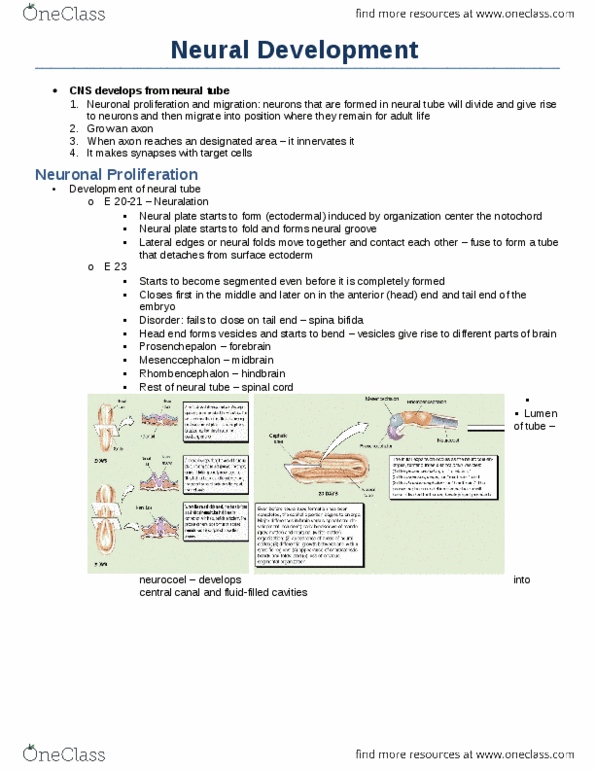
1. Embryologic brain origins:
1) Neural plate: from ectoderm thickening (3rd week)
2) Neural plate invaginates
a. =Groove flanked by crests: neural folds
3) Neural folds fuse, separate from ectoderm= neural tube (4th week)
4) Neurocoel: neural tube central cavity:
a. Entire CNS derives from neurocoel
5) Neural crest: ectoderm pinched off, just lateral to neural tube
a. Forms somites (bulges) either side of neural tube
b. Somites = dermis, muscles, skeleton
6) Neural tube differentiates into CNS:
a. Anterior enlarges = primary brain vesicles
i. Fluid filled cavities w/in neural tube
ii. Proencephalon (forebrain)
iii. Mesencephalon (midbrain)
iv. Rhombencephalon (hindbrain)
b. Rest of neural tube = spinal cord
7) Primary vesicles → secondary vesicles (5th week)
a. Proencephalon → telencephalon + diencephalon
b. Mesencephalon (no change)
c. Rhombencephalon → metencephalon + myelencephalon
8) Secondary vesicles → adult brain structures
a. Telencephalon: cerebrum
b. Diencephalon: epithalamus, thalamus, hypothalamus
c. Mesencephalon: midbrain
d. Metencephalon: pons, cerebellum
e. Myelencephalon: medulla oblongata
9) Central cavity forms 4 ventricles of brain
2. What primary germ layer gives rise to nervous tissue? Ectoderm
3. Brain structures associated with primary brain vesicles:
1) Proencephalon: cerebrum, epithalamus, hypothalamus, thalamus
2) Mesencephalon: midbrain
3) Rhombencephalon: medulla, pons, cerebellum
find more resources at oneclass.com
find more resources at oneclass.com

4-6. Two common types of neural tube defects:
1) Anencephaly: failure to close anterior neural tube
a. =Skull/brain not completed, exposed to air =fatal
b. Prevented by dietary supplement: Folic acid
2) Spina Bifida: failure to close posterior (caudal) neural tube
a. Cleft spine
b. Closed: hairy patch, birth defects
c. Menigocele: spinal cord protrudes through opening
d. Myelomeningocele: bones of spine don’t form completely
=incomplete spinal canal
7. Spina bifida newborn assessment findings:
A. Urinary/bowel issues
B. Paralysis
C. Abnormal hair tuft on spine
D. Collection fat
E. Dimple, birthmark
F. Seizures
G. Muscle weakness
8. Structural outcomes of brain developing w/in restricted space:
A. Midbrain & cervical flexures:
a. Brain bends forward during growth
B. Posterior & lateral growth cerebral hemispheres:
a. Wrap around top of brainstem
C. Surface convolutions: (gyri & sulci)
a. High SA= more brain cells
9. Ventricles of brain & their connections:
• Continuous w/ one another & spinal cord
1) Lateral ventricles: C-shaped chambers separated by septum pellucidum
*Connects to third ventricle via: interventricular foramen*
2) Third ventricle: surrounded by hypo/epi/thalamus
*Connects to fourth ventricle via: cerebral aqueduct*
3) Fourth ventricle: behind pons & medulla
*Connects to spinal cord & subarachnoid space via: three openings…*
1&2: Lateral apertures: in side walls
3. Median aperture: in roof of 4th ventricle
find more resources at oneclass.com
find more resources at oneclass.com

10. Fissures/sulci & lobes of cerebrum:
1) Central sulcus: separates frontal & parietal
2) Parieto-occipital sulcus: separates parietal & occipital
3) Lateral sulcus: separates temporal from parietal & frontal
4) Longitudinal fissure: separates cerebral hemispheres
5) Transverse cerebral fissure: separates cerebrum & cerebellum
*Insula: covered by temporal, parietal, frontal lobes
11-12. Function & Location of following cortical areas:
1) Primary motor cortex:
a. Location: frontal lobe
b. Pyramidal cells
c. Controls voluntary contractions of specific skeletal muscles (on
opposite sides of body)
2) Premotor cortex:
a. Location: frontal lobe
b. Planning movement, controlling learned/skilled movement
3) Broca’s area:
a. Motor speech area
b. Location: frontal lobe…
c. Translates thoughts → motor patterns of speech
4) Frontal eye field:
a. Location: frontal lobe
b. Voluntary eye scanning movement
5) Primary auditory cortex:
a. Location: temporal lobe
b. Receives input from cochlea
c. =Detects: pitch, rhythm, volume
6) Auditory association area:
a. Location: inf/post to primary auditory cortex (temporal)
b. Perception/interpretation of auditory input
7) Prefrontal cortex:
a. Location: frontal lobes
b. Very complicated processing: intellect, complex learning
c. Develops in adolescence
8) General interpretative area:
a. Wernicke’s area & gnostic area (frontal)
b. Pattern recognition, language interpretation, articulation
c. Damage= people don’t understand what they are reading/hearing
9) Primary somatosensory cortex:
a. Location: parietal lobe
b. Receive stimuli from contralateral body somatic sensory receptors
i. Ex: skin, joints, skeletal muscle
find more resources at oneclass.com
find more resources at oneclass.com