BM 1041:03 Lecture Notes - Lecture 26: Carpal Tunnel Syndrome, Carpal Tunnel, Cubital Fossa

23 May 2018
School
Department
Course
Professor

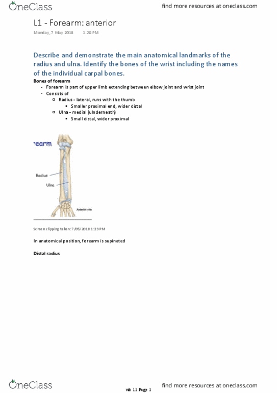
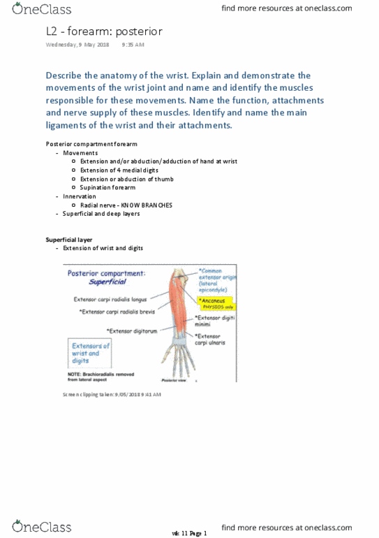
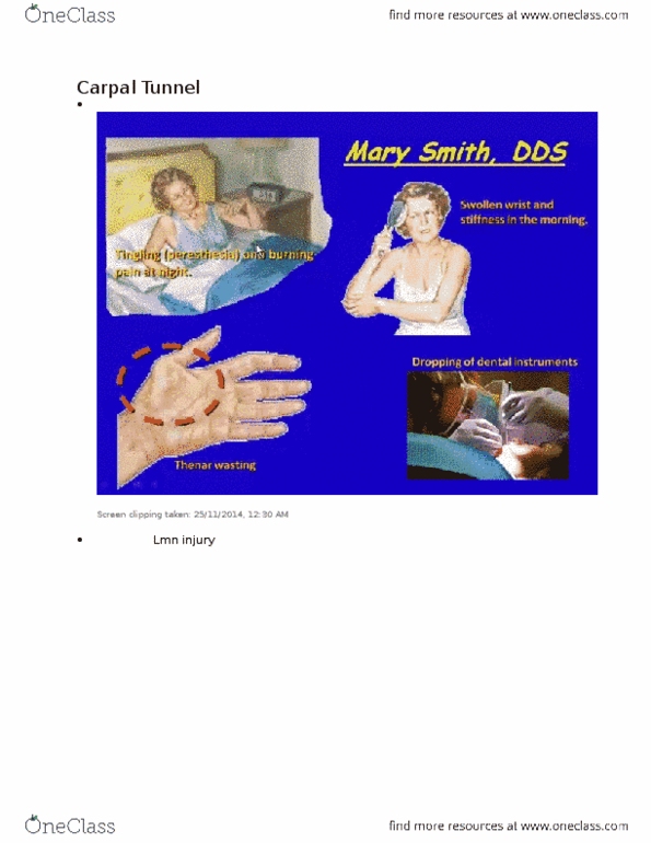
Describe the boundaries and contents of the carpal tunnel and the
anatomical and functional significance of carpal tunnel syndrome.
Key transition areas
Screen clipping taken: 9/05/2018 10:14 AM
Cubital fossa (from last week)
-
Screen clipping taken: 9/05/2018 10:14 AM
Distally structures pass between forearm and hand through or anterior to carpal tunnel
○
EXCEPT radial artery, which passes dorsally around wrist
○
Carpal bones DO NOT lie in a flat plane, instead form an arch
○
Roof/anterior wall of tunnel = flexor retinaculum ligament, spanning distance between
medial and lateral sides of base
○
Lateral side of base = tubercles of scaphoid and trapezius
○
Medial side = pisiform, hook of hamate
○
Carpal tunnel
-
L3 - wrist and carpal tunnel
Wednesday, 9 May 2018
10:13 AM
wk 11 Page 1
Document Summary
Describe the boundaries and contents of the carpal tunnel and the anatomical and functional significance of carpal tunnel syndrome. Distally structures pass between forearm and hand through or anterior to carpal tunnel. Except radial artery, which passes dorsally around wrist. Carpal bones do not lie in a flat plane, instead form an arch. Roof/anterior wall of tunnel = flexor retinaculum ligament, spanning distance between medial and lateral sides of base. Lateral side of base = tubercles of scaphoid and trapezius. Medial side = pisiform, hook of hamate wk 11 page 1. 4 tendons of flexor digitorum superficialis single tendon of flexor pollicis longus. Tendons of fdp and fds are surrounded by single synovial sheath. Ligament holds tendons to bony plane at the wrist. Caused by pressure on median nerve within carpal tunnel. May be direct effect of increased pressure on median nerve caused by overuse, swelling of tendons, tendon sheaths and cysts arising from carpal joints.