ANAT20006 Lecture Notes - Lecture 29: Peritoneal Cavity, Peritoneum, Gastric Mucosa

12 Jun 2018
School
Department
Course
Professor

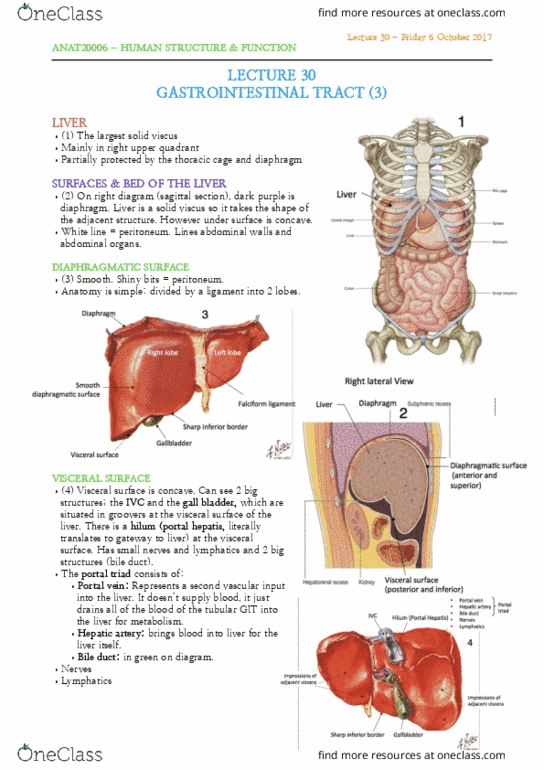
LECTURE 29
GASTROINTESTINAL TRACT (2)
ABDOMINAL VISCERA
& QUADRANTS
•(1) Divided into 4 quadrants
for ease of identification.
PERITONEUM
•(2) Peritoneum: Serous
membrane lining abdominal walls
& some of the organs/viscera.
Parietal (walls) vs visceral
(abdominal organ surface).
•The abdomen is actually kidney
shaped (not an oval) due to the
lumbar vertebrae behind it
pushing on it.
•The kidney is considered to be
part of the wall structure
(posterior abdominal wall) and is
therefore considered to have
parietal peritoneum.
•Peritoneal cavity is a
potential space
between the peritoneal
membranes with a few
mLs of serous fluid.
•Abdominal viscera:
Intraperitoneal
(within peritoneal
cavity, have full
coat of peritoneal
membrane and
mesentery) vs
Retroperitoneal
(outside peritoneal
cavity, have fixed position and no mesentery).
•Mesentery
•On right diagram: white lines represent peritoneal. Lines
some aspects of pelvic viscera. Thus peritoneum is not only in
the abdomen; can extend to pelvis.
OESOPHAGUS
•(3) Muscular tube beginning at level C6 (same as trachea).
Descends at the midline and at level T10 it goes into the
diaphragm and passes down to the abdomen to enter the
stomach from the right hand side. It is the beginning of the
GIT but the majority is in the thorax, not the abdomen.
•Narrowing at beginning, middle and end as it is a muscular
tube. Muscular part of diaphragm surrounding oesophagus at level
T10 forms a functional sphincter.
•At the T4 T5 level, the trachea gives rise to the bronchi, and the
Lecture 29 - Wednesday 4 October 2017
ANAT20006 - HUMAN STRUCTURE & FUNCTION

aorta begins to arch upwards toward the left and
downwards. So at T4 T5 the left main bronchus and the
aortic arch externally compress the oesophagus, causing a
constriction/narrowing.
•(4) 2 complete layers of muscular walls. External/
longitudinal layer and inner/circular layer.
•Once the oesophagus enters the stomach, the oesophageal
mucosa changes sharply to become gastric mucosa.
There is a small visible line that we can see, the
oesophagogastric junction.
STOMACH
ANTERIOR VIEW
•(5) Left upper quadrant.
•Intraperitoneal
•Features include:
•1. Two orifices
•2. Two curvatures
•3. Two surfaces
•4. Four parts
•Majority of GIT is tubular but stomach is
dilated, usually a rough J shape. It is a hollow
viscus so it is divided into 4 parts. The beginning
(entry) is the cardiac part of the stomach.
The top part above the opening is the
fundus. Middle/majority section is the body.
The bottom is the pyloric part, where it
begins to narrow. This has 2 further parts:
the pyloric entry (first bit after body) and
then the pyloric canal (the last bit).
•There is a cardiac opening
next to the cardiac part;
opening to the
oesophagus. The pyloric
orifice opens onto the
intestines. This is an
example of a true
anatomical sphincter, the
pyloric sphincter.
INTERNAL VIEW
•(6) Rugae is gastric
mucosa folds.
Lecture 29 - Wednesday 4 October 2017
ANAT20006 - HUMAN STRUCTURE & FUNCTION
Document Summary
& quadrants: (1) divided into 4 quadrants for ease of identification. Peritoneum: (2) peritoneum: serous membrane lining abdominal walls. Intraperitoneal (within peritoneal cavity, have full coat of peritoneal membrane and mesentery) vs. Retroperitoneal (outside peritoneal cavity, have fixed position and no mesentery): mesentery, on right diagram: white lines represent peritoneal. Thus peritoneum is not only in the abdomen; can extend to pelvis. Oesophagus: (3) muscular tube beginning at level c6 (same as trachea). Descends at the midline and at level t10 it goes into the diaphragm and passes down to the abdomen to enter the stomach from the right hand side. Git but the majority is in the thorax, not the abdomen: narrowing at beginning, middle and end as it is a muscular tube. Muscular part of diaphragm surrounding oesophagus at level. T10 forms a functional sphincter: at the t4 t5 level, the trachea gives rise to the bronchi, and the.