ANAT20006 Lecture Notes - Lecture 6: Limb Bud, Morning Sickness, Neural Crest

13 Aug 2018
School
Department
Course
Professor
Document Summary
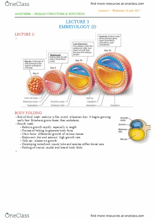
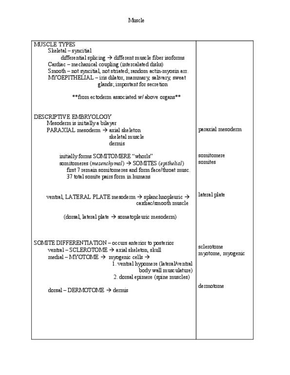
Fate of mesoderm: prof colin anderson c. anderson@unimelb. edu. au. Divided into: paraxial mesoderm (medial, intermediate mesoderm, lateral mesoderm. Middle brown layers and can be divided into 3 parts from the notochord. Ventrolateral body wall (connective tissue (incl dermis), not muscle) Swellings (somitomere) appear progressively down length of paraxial mesoderm. At the 20 somitomere stage, the eighth pair of somitomere becomes an independent pair of somites. Starts when the medial strip of the mesoderm starts to change by forming a series of swelling down the length called somitomeres swellings (somitomeres) appear progressively down length of paraxial mesoderm. Blue is the neural tube with the notochord underneath. The swelling are thickening in the continuous sheath of the mesoderm. You turn these lumps into separate discrete entities and become somites. The first somites occur in the position of the 8th somitomere counting down form the cranial end of the animal. The first ones to break free from the surrounding mesoderm.