PSYC10003 Lecture 9: PSYC10003 9. The Sensorimotor System


9. The Sensorimotor System
Two patients with Motor Impairments after Stroke
EH:
• 68 year old man
• ake up ouldt oe left ar & leg
• admitted to hospital, neurologist guessed stroke
• neurological examination showed EH could not make
voluntary movements with his left limbs
o but preserved stretch reflexes
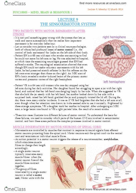
• brain scan revealed stroke-induced lesion of primary
motor cortex and premotor cortex RH
VR:
• 64 year old woman
• one day stopped using left arm during daily activities
• found struggling open jar with right hand with left
hand dangling limply
• surprised when noticed and started using left arm
• many similar incidents during day as if VR forgot she
had left arm
• while attention drawn to it – could use it normally
• i M‘I, large lesio foud i V‘s right parietal loe fro stroke
Motor control: control of body
Two cases illustrate two different failures of motor control.
Muscle Innervation by Motor Neurons of Spinal Cord
• movements controlled by muscles that contract in response
to neural signals from efferent motor neurons projecting
from spinal cord
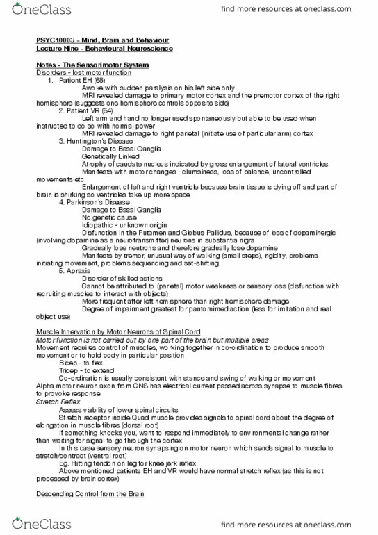
• motor neurons exit spinal cord from ventral root and
terminate on individual muscle fibres
• AP in motor neuron triggers release of neurotransmitter
acetylcholine, stimulating muscle fibres to change their
length accordingly
• Single motor neuron typically innervates many muscle
fibres
• When motor neuron fires all the muscle fibres contract
together
• Group of fibres innervated by single motor neuron is a
motor unit
o Motor units with fewest muscle fibres (eg. In
face/hands) permit greatest degree of selective motor control
find more resources at oneclass.com
find more resources at oneclass.com

2
• Many skeletal muscles fall into two categories:
o Flexors: at to ed or fle a joint
o Extensors: act to straighten a joint
o These categories of muscle often act antagonistically (ie.
In opposition, eg. Biceps/triceps)
Stretch Reflex
• Spinal motor neurons receive input from variety of sources
o A source is the sensory receptors located within muscles themselves
• Activity of skeletal muscles is monitored by receptors called muscle spindles
o Provide info to CNS regarding muscle length
• When muscle unexpectedly stretched (eg. When hammer taps beneath patella)
o Muscle spindles convey info
back to spinal cord via dorsal
roots
o Axons of spindle afferent
neurons synaps directly with
motor neurons
o → irease atiit i order to
return muscle to original length
o results in brisk contraction of
quadriceps muscles, causing
lower leg to extend
o ↑ iruit fors siple reflex
arc
• functional significance: compensate for any perturbation by external forces and thus maintaining
intended position of body
• eg. When someone bumps into you/brushes your arm while carrying coffee, stretch reflex
compensates automatically and prevents falling/spilling drink
• EH and VR both had strong patellar tendon reflexes, indication motor and sensory neurons of the
spinal cord remained intact
Preserved walking following Spinal Cord Resection
• Motor neurons in spinal cord capable of triggering complex movements of various muscle groups
without any controlling signals from brain
• Illustrated in cats, spinal cord is surgically sectioned at a point just above where spinal nerves
subserving hind legs are located
o Effectively disconnects lower motor neurons for hind legs from the brain
• Despite spinal cord section, cats still able to walk normally on treadmill
o Showing normal extensor and flexor movements of hind legs
find more resources at oneclass.com
find more resources at oneclass.com

3
• dramatic illustration of hierarchical
organization of motor system
o motor and sensory neurons
within spinal cord are able to
control all complex patterns of
muscle contraction required
for walking without
instruction from brain
o leaves brain free to control
more demanding aspects of
motor control
o eg. Determining precisely
when initiating particular actions
Descending Control from the Brain
• most purposeful actions are initiated and controlled voluntarily & actions such actions depend upon
signals generated by brain that are conveyed to the muscles via spinal cord
• eg. Lecture 2 hot casserole dish excitation is counteracted by inhibitory input from primary motor
cortex in brain.
o The axons that descend from primary motor cortex through spinal cord form inhibitory
synapses with lower motor neurons
o These inhibitory synapses can prevent muscle contraction from occurring by blocking AP
o Similarly, excitatory inputs from brain can trigger AP in lower motor neurons and initiate
movements
• EH unable to make voluntary movements with left limbs
o Signals from primary motor cortex are evidently needed for voluntary movements with left
limbs
• VR able to make normal limb movements when prompted but cannot do so spontaneously
o Parietal lesion ∴ seems to have affected capacity to initiate movements internally
Hierarchical Control in the Sensorimotor System
• Human sensorimotor system: commands are issued in a top-down manner
• Association areas (prefrontal cortex & parietal cortex) act as president, specifying goals rather than
specific plans of action; not involved in details
o Leaves highest levels of control free to perform most complex functions
• Secondary motor cortex (the premotor and supplementary motor areas) involved in programming
specific patterns of movements
• Primary motor cortex: point of departure from which sensorimotor signals from brain are conveyed
to brainstem and spinal cord
• the hierarchical organization involves both top-down and bottom-up
• if problem arises with one of workers, this is conveyed back up chain of command to higher levels
whose responsibility it is to resolve any problems, just as sensory feedback from the muscles and
tendons is monitored by CNS in case adjustments are required
find more resources at oneclass.com
find more resources at oneclass.com
Document Summary
Two cases illustrate two different failures of motor control. Preserved walking following spinal cord resection: motor neurons in spinal cord capable of triggering complex movements of various muscle groups without any controlling signals from brain. Cortical regions involved in motor function: at cortical level, there are several key structures involved in sensorimotor control, prefrontal & parietal cortex acts as president, vr patients fails to spontaneously move left arm & hand. In a situation: neurological disorders of hu(cid:374)ti(cid:374)gto(cid:374)"s chorea and parki(cid:374)so(cid:374)"s disease are chracterised by dysfunction of basal ganglia, cerebellum: timing, prediction of sensory consequences of movement, basal ganglia: initiation, sequencing, set-shifting. Many were executed, believed to be possessed by evil spirits. Pd patients may have problems varying the force required to perform limb movements, producing series of small bursts of agonist and antagonist muscles rather than scaling a single agonist burst to reach goal.