PSYC10003 Lecture Notes - Lecture 5: Primary Motor Cortex, Temporal Lobe, Abdominal Cavity

6 Jun 2018
School
Department
Course
Professor


PSYC10003 – MIND, BRAIN, & BEHAVIOUR 1
BEHAVIOURAL NEUROSCIENCE
Lecture 5 (Week 2 . 2): Structure & Function of the Human Nervous System – Part 1
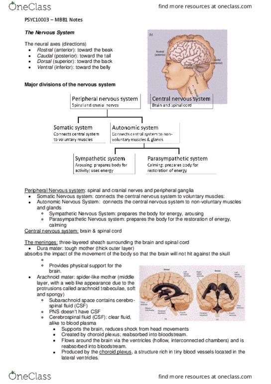
The neural axes: an imaginary line drawn through the spinal cord toward the front of the brain. Used to
locate structures within the nervous system.
• Rostral (anterior): ‘toward the beak’
• Caudal (posterior): ‘toward the tail’
• Dorsal (superior): ‘toward the back’
• Ventral (inferior): ‘toward the belly’
o Lateral: toward the side
o Medial: toward the midline
➢ Ipsilateral: structures on the same side of the body
➢ Contralateral: structures on the opposite side of the body
Major Divisions of the Nervous System:
• Central Nervous System (CNS): includes the brain & spinal cord
• Peripheral Nervous System (PNS): includes the cranial nerves, spinal nerves, & peripheral ganglia
• Somatic Nervous System: connects the CNS to voluntary muscles
• Autonomic Nervous System: connects the CNS to non-voluntary muscles & glands
• Sympathetic Nervous System: arousing; prepares the body for activity,
expends energy
• Parasympathetic system: calming; prepares the body for restoration of
energy
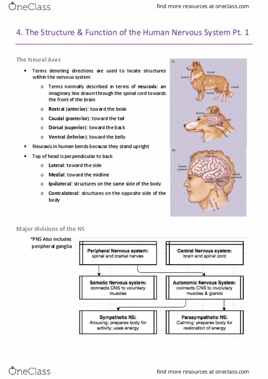
The Menings: NS’s protective sheaths of connective tissue. CNS has all, PNS: dura
mater & pia mater (fuse into 1 layer covering spinal nerves & peripheral ganglia)
• Dura mater (‘tough mother’): the thick outer layer
• Arachnoid mater (‘spider-like mother’): middle layer, weblike appearance due to the protrusions
(arachnoid trabeculae), is soft & spongy
• Pia mater (‘pious mother’): clear, delicate inner layer, follows folds of brain tissue
• Space between the arachnoid & pia mater: subarachnoid space.
Contains blood vessels, & fluid that bathes the brain & spinal cord
Cerebrospinal Fluid: fluid that supports & protects the brain & spinal cord
• Found in the subarachnoid space, around the outside of the brain &
spinal cord, & also in the hollow, interconnected chambers inside the brain (ventricles)
• Produced by choroid plexus in lateral ventricles (1 per hemisphere) → flows down to third ventricle
→ flows through cerebral aqueduct to fourth ventricle → exits via a set of openings into the
subarachnoid space → reabsorbed into the bloodstream via the arachnoid villae
Ventricle system: set of linked, fluid-filled chambers (ventricles).
• There’s 2 lateral ventricles (1 per hemisphere), linked centrally
with the third ventricle, which connects to the forth ventricle
immediately beneath the cerebellum via the cerebral aqueduct
• Hydrocephalus: occurs when the flow of CSF is blocked,
causing CSF to accumulate within the ventricles, raising the
pressure inside the skull, potentially leading to brain damage.
find more resources at oneclass.com
find more resources at oneclass.com