BIOM3002 Lecture Notes - Lecture 4: Stratified Squamous Epithelium, Muscularis Mucosae, Lamina Propria

26 May 2018
School
Department
Course
Professor

HISTOLOGY
HISTOLOGY LECTURE FOUR
DIGESTIVE SYSTEM FUNCTIONS:
1. Ingestion
2. Mechanical breakdown
3. Secretion [saliva, gastric juice, bile, pancreatic juice]
4. Digestion [chemical treatment] food [enzymes, sectretions]
5. Transportation
6. Absorption nutrients: non-digested nutrients/micronutrients
I.e. water/minerals/vitamins; and digested
nutrients/macronutrients I.e. AA's, fatty acids, glucose
7. Excretion
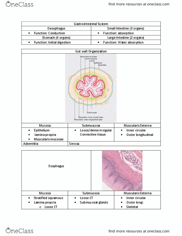
4 x layers gut tube wall
1. MUCOSA
• In contact with food
• Varies most
• Mucosal epithelium
• Mucosal connective tissue [lamina propria]
• Muscularis mucosae [churning – GIT only]
2. SUBMUCOSA
3. MUSCULARIS EXTERNA [outer longitudinal, inner circular
layer]
4. SEROSA/ADVENTITIA [epithelium, CT]
4 x functional types of mucosa
1. PROTECTIVE: stratified squamous epithelium, oral cavity,
oesophagus, anal canal
2. SECRETORY: gastric glands, stomach [acids and enzymes and
mucous]
3. ABSORPTIVE: columnar epithelium, villus, small intestine
4. ABSORPTIVE/PROTECTIVE: colon, columnar epithelium, tubular
glands
ABRUPT hanges occur from one type of mucosa to another – think
about implications
Loose vs dense connective tissue:
L: lamina propria – loose areolar CT, strongly allied with the
immune system [dark dots = lymphocytes]
D: submucosa contains more dense irregular CT; some regions
more dense than others
Skeletal vs smooth muscle:
Smooth: irregular arrangement, single nuclei, 'wring-out'
Skeletal: top and bottom GIT, one direction contraction,
multinucleated cells, lienar
find more resources at oneclass.com
find more resources at oneclass.com

HISTOLOGY
GALT: GUT-ASSOCIATED LYMPHOID TISSUE
• MALT [mucosa-associated lymphoid tissue] is constitutively
present unlike respiratory system
• Accumulations of lymphocytes in lamina propria
• Accumulated lymphocytes organised into nodules/follicles
OESOPHAGUS:
• Protective epithelium
• Scattered lymphocytes
• Muscularis externa = circular muscle and longitudinal muscle
• Top 1/3 = skeletal muscle; gradual transition as peristalsis
takes over
• Submucosa: mucous-secreting glands [muscularis mucosae and
glandular tissue]
• Local nerve plexes; ANS control[myenteric plexus]
• No epithelium outside [adventitia]
Gastro-oesophageal junction: muscularis externa layer thickens
at sphincter
Stomach muscularis external and overview:
• Mucosa and gastric glands
• Submucosa
• Oblique muscle layer
• Inner muscle layer
• Outer muscle layer
Gastric pits and glands; how do we control secretion?
• Invaginated simple epithelium
• Between glands = CT [lamina propria]
• Endocrine secreting cells
Gastric pits [foveolae]
Gastric glands
Chief cells secrete enzymes in inactive form – pass parietal
cells
Parietal cells secrete HCl [protective against pathogens;
cleaves enzymes into active form]
Rest of gastric pit secretes mucous
Stomach lining rapidly regenerating
Threads of muscularis mucosae [helps secretion] between
glands
Colour mucous-secreting cells = quite pale as PAS stains
mucous
find more resources at oneclass.com
find more resources at oneclass.com
Document Summary
Digestive system functions: ingestion, mechanical breakdown, secretion [saliva, gastric juice, bile, pancreatic juice, digestion [chemical treatment] food [enzymes, sectretions, transportation, absorption nutrients: non-digested nutrients/micronutrients. 4 x layers gut tube wall: mucosa, in contact with food, varies most, mucosal epithelium, mucosal connective tissue [lamina propria, muscularis mucosae [churning git only, submucosa, muscularis externa [outer longitudinal, inner circular layer, serosa/adventitia [epithelium, ct] 4 x functional types of mucosa: protective: stratified squamous epithelium, oral cavity, oesophagus, anal canal, secretory: gastric glands, stomach [acids and enzymes and mucous, absorptive: columnar epithelium, villus, small intestine, absorptive/protective: colon, columnar epithelium, tubular glands. Abrupt hanges occur from one type of mucosa to another think about implications. L: lamina propria loose areolar ct, strongly allied with the immune system [dark dots = lymphocytes] D: submucosa contains more dense irregular ct; some regions more dense than others. Skeletal: top and bottom git, one direction contraction, multinucleated cells, lienar.