6529 Lecture Notes - Lecture 7: Superior Vena Cava, Pulmonary Artery, Pulmonary Vein

19 Jun 2018
School
Department
Course
Professor

Cardiovascular System
Heart Anatomy
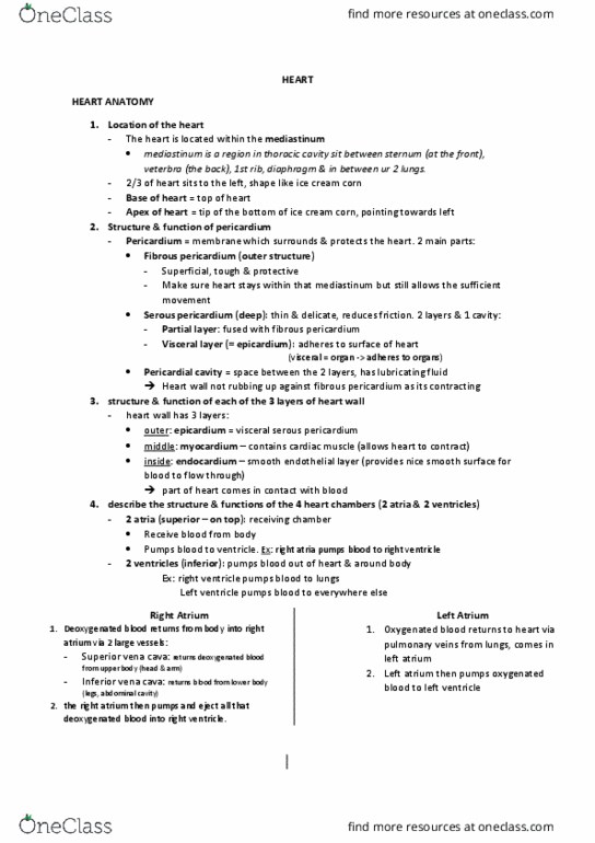
Heart is located in the thorax – called the mediastinum ( medi – medial , stinum =
sternum region )
Heart has its own casing – called the pericardium – it holds the heart in place.
Pericardium – has 2 layers
Serous pericardium – which has 2 layers
- Visceral = means it is closest to the organ
- Parietal is the outer most layer.
Fibrous pericardium
- Is a tough connective tissue that protects the heart , and anchors the heart to surrounding
structures and it also helps prevents overfilling of the heart with blood.
Wall of the heart - 3 layers
-Epicardium = is the layer
of serious pericardium
-Myocardium – does the
contractions for heart
-Endocardium = is the
inner heart lining and it is
continuous with exiting
vessels. Can become
inflamed
find more resources at oneclass.com
find more resources at oneclass.com

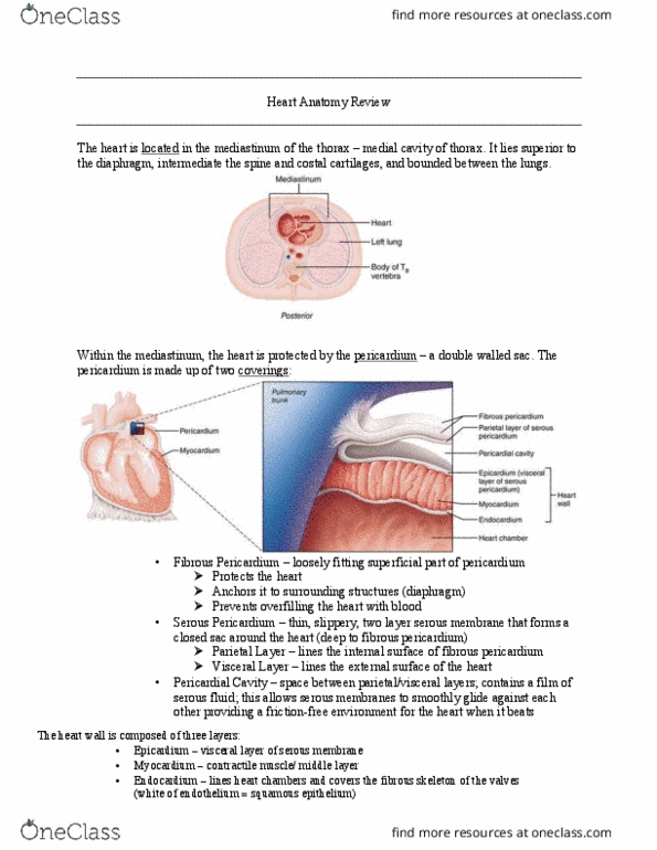
Chambers of the
heart
Anterior is
rounded
Posterior is flat
Blue –
deoxygenated
Red – oxygenated
Pathway of blood
find more resources at oneclass.com
find more resources at oneclass.com

- Superior and inferior vena cava are the main veins that receive blood from the body. The
superior vena cava drains the head and arms, and the inferior vena cava drains the lower
bod
- right atrium- receives blood form the body via the vena cava. The atria are on the top in the
heart.
- Tricuspid Valve- The blood then passes through the right atrioventiricular valve (tricuspid),
which is forced shut when the ventricles contract, preventing blood from re-entering the
atrium.
- Right vemntricle - The blood goes into the right ventricle (note that it has a thinner wall; it
only pumps to lungs). The ventricles are on the bottom of the heart.
Out the pulmonary trunk to the lungs
- pulmonary artery/trunk is the main artery taking deoxygenated blood the lungs. Blood goes
- to the right and left lungs, where capillaries are in close contact with the thin-walled alveoli
so the blood can release CO2 and pick up O2.
From lungs - Recive oxygenated blood back to the heart via pukmonary veins
Veins = return blood to the heart
Arterys = take blood away frm the heart.
- pulmonary vein carries oxygenated blood back into the heart
- left atrium receives oxygenated blood from the lungs. The blood passes through
- the left atrioventricular valve (bicuspid or mitral valve).
- The blood enters the left ventricle. Note the thickened wall; the left ventricle must pump
blood throughout the entire body
- The blood passes through the left semilunar valve at the beginning of the aorta. The aorta is
the main artery to the body. One of the first arteries to branch off is the coronary artery,
which supplies blood to the heart muscle itself so it can pump
From the capillaries in the heart muscle, the blood flows back through the coronary vein, which
lies on top of the artery.
- The aorta divides into arteries to distribute blood to the body. Small arteries are called
arterioles. The smallest vessels are the capillaries. The capillaries feed into the
venules, which feed into veins which feed into the superior and inferior vena cava
(and back to step 1).
find more resources at oneclass.com
find more resources at oneclass.com
Document Summary
Heart is located in the thorax called the mediastinum ( medi medial , stinum = sternum region ) Heart has its own casing called the pericardium it holds the heart in place. Visceral = means it is closest to the organ. Is a tough connective tissue that protects the heart , and anchors the heart to surrounding structures and it also helps prevents overfilling of the heart with blood. Epicardium = is the layer of serious pericardium. Myocardium does the contractions for heart. Endocardium = is the inner heart lining and it is continuous with exiting vessels. Superior and inferior vena cava are the main veins that receive blood from the body. The superior vena cava drains the head and arms, and the inferior vena cava drains the lower bod right atrium- receives blood form the body via the vena cava. The atria are on the top in the heart.