ANHB1102 Lecture Notes - Lecture 11: Respiratory Epithelium, Pleural Cavity, Mucous Membrane

25 May 2018
School
Course
Professor

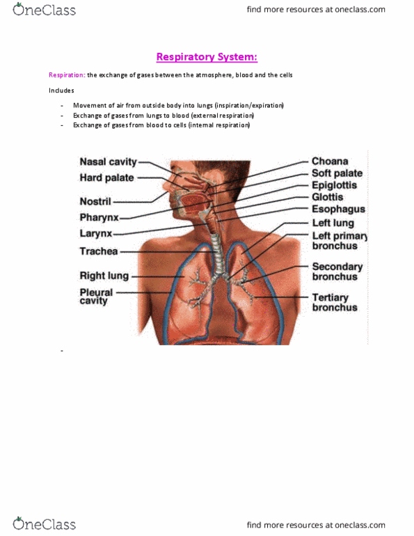
Respiratory Notes:
-Respiration: the exchange of gases bet2ween the atmosphere, blood and the cells
-movement of air from outside the body into the lungs (inspiration/expiration)
-exchange of gases from the lungs to the blood (external respiration)
-exchange of gases from the blood to the cells (internal respiration)
Gross Anatomy of Respiratory System:
Functions of Respiratory System:
-Move air into and from the body (for O2 and CO2 exchange)
-Non-respiratory Functions:
-Provide nonspecific defences against pathogens
-Homeostasis of body fluids (especially pH)
-Vocal communication
-Olfaction
Development of Respiratory System:
-4 Weeks: Buds of foregut endoderm form pleural cavity
-5 Weeks: Buds continue to grow and branch (secondary bronchi)
-8 Weeks: Mesoderm forms cartilages, muscle, connective tissue supports
-24 Weeks: alveoli developed sufficiently for survival
-24-28 Weeks: surfactant production begins and production increases in final two months in utero
(prevents alveoli cell walls from sticking together)
-Birth/First Breath: first gasp takes supreme effort, and lungs are never completely empty again

Respiratory Mucosa and Epithelium:
-Mucous membrane = respiratory epithelium + underlying connective tissue
-Respiratory epithelium = pseudo stratified ciliated columnar epithelium with goblet cells
-Connective tissue with mucous and serous glands + blood sinusoids
-Mucosa lines entire tract down to respiratory bronchioles
Gross Anatomy and Function of Respiratory Tract:
Nasal Cavity: Functions:
-Modifies air entering respiratory tract
-Warms - blood sinusoids (very vascular)
-Moistens - glands (e.g mucous glands)
-Cleans - cilia + mucous and hairs
-Olfaction: sense of smell
-Specialised epithelium detect smell -> CNS
Pharynx & Larynx:
-Swallowing: epiglottis folds
down onto larynx to prevent
food and liquid entering the
trachea and directs it into the
esophagus

Larynx:
-Comprised of
cartilages, ligaments
and muscles
-Functions:
-Protects the airway
(cough reflex)
-Phonation
(production of
speech sounds)
Trachea:
-Leads from larynx to primary
bronchi
-Lined with respiratory epithelium
-C shaped cartilages support wall
-Rings are not complete to allow
for stretch of esophagus when
food travels through it
-Receptors detect temperature and
pressure (e.g if food enters)
Lungs:
-Left lung has only 2 lobes to allow for heart
-Lobes are completely separate from each other
-Right lung is shorter due to diaphragm and liver
Pleura:
-Visceral pleura covers the surface of the lungs and
extends into the fissures
-Parietal pleura adheres to the mediastinum, inner surface
of rib cage and superior surface of diaphragm
-Pleural cavity contains pleural fluid and is a potential
space (normally no room between membranes)
-Functions:
1. Reduction of friction: pleural fluid acts as lubricant
and enables lungs to expand and contract with
minimal friction
Document Summary
Respiration: the exchange of gases bet2ween the atmosphere, blood and the cells. Movement of air from outside the body into the lungs (inspiration/expiration) Exchange of gases from the lungs to the blood (external respiration) Exchange of gases from the blood to the cells (internal respiration) Move air into and from the body (for o2 and co2 exchange) 4 weeks: buds of foregut endoderm form pleural cavity. 5 weeks: buds continue to grow and branch (secondary bronchi) 8 weeks: mesoderm forms cartilages, muscle, connective tissue supports. 24 weeks: alveoli developed suf ciently for survival. 24-28 weeks: surfactant production begins and production increases in nal two months in utero (prevents alveoli cell walls from sticking together) Birth/first breath: rst gasp takes supreme effort, and lungs are never completely empty again. Mucous membrane = respiratory epithelium + underlying connective tissue. Respiratory epithelium = pseudo strati ed ciliated columnar epithelium with goblet cells. Connective tissue with mucous and serous glands + blood sinusoids.